Virtual reconstruction of the vertebrate skeleton,
used for visualization and functional analysis of the object, makes it possible
to extract more complete information about the biological role of the integral
structure and the adaptive meaning of its transformations, which represents a
significant addition to classical morphometric research methods. It is known
that the leading factor determining bone tissue formation is mechanical
loading, which changes during onto and phylogenesis [1, 2], indicating
that adaptive “deformation” of bone occurs at sites of
greatest stress. In this regard, the question of the localization and
distribution of stresses and strains in an object of given shape remains open.
One of the modern methodological approaches that allows us to approach the
solution of this problem is the determination of the stress–strain state of
bone tissue under the action of external forces. The most widely used
mathematical method for strength analysis of structures is the finite element
method, which makes it possible to identify stresses and strains in an object
of any shape and geometry under specified loads and mechanical characteristics
of the object under study. To date, the use of this approach has made it
possible to perform virtual reconstructions of the skeleton of some species of
large mammals and extinct reptiles, as well as to analyze aspects of their
functional and evolutionary morphology [3, 17]. Based on the experience of
previous researchers, we have developed and tested a method for step by step
assembly of tomographic sections and the creation of a virtual construction of
the fish skeleton, which allows obtaining primary data on the mechanical
behavior of bone tissue in this group of vertebrates [18, 19]. The present work
is a continuation of the initiated research and is devoted to the application
of the finite element method for the analysis of adaptive transformations of
the fish jaw apparatus.
The main stages of modeling the FEM of the fish viscerocranium were presented in detail in previous works
[18, 19]. In the present study, in order to fully demonstrate the application
of FEM for studying adaptive transformations of the fish jaw apparatus, we
present an algorithm and a brief description of the main stages of model
creation for testing (Fig. 1).
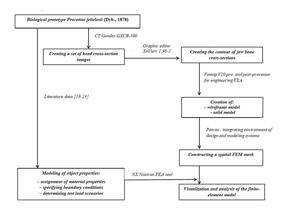
Fig. 1. Algorithm for creating and analyzing the FEM of the fish jaw apparatus [19].
Creation of the FEM of the fish jaw apparatus.
The biological prototype of the model being created
was a collection specimen of the Baikal endemic ichthyofauna – Procottus jeitelesii
(Dyb., 1878). Scanning of the object was performed using a Gendex
GXCB 50 cone beam volumetric tomography unit
(https://angarsk-stomklinika.ru) in a vertical position, from the symphysis to
the posterior edge of the operculum. The image frame parameters were 216×216 mm
with a slice step of 0.4 mm. Visualization of the results of
layer‑by‑layer scanning of the fish skull was carried out using
iCATVision 1.8 software for visualization of computed
tomography data of the maxillofacial region [20]. The source materials were
raster images in JPEG format with a size of 312×312 pixels (Fig. 2 a).
A total of 106 section images of the head were obtained and analyzed.
Analysis of the obtained sections was performed using
a color RGB model in the XnView 1.98.2 image viewing
and editing software [21]. The contours of the object were created by
vectorizing points in the color gradient at the boundary
between the background and the bone section (Fig. 2 a). For subsequent data
processing in engineering analysis programs, the obtained section contour
images were converted into DXF and NEU formats.
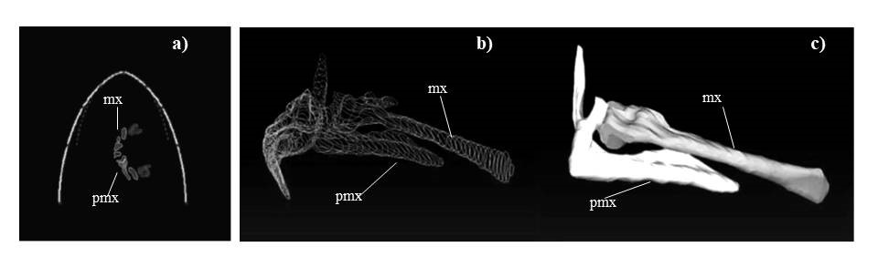
Fig. 2. Creation of a solid model of the maxillary bones of the fish (P. jeitelesii): a) bone contours in the raster image of CT
sections of the head; b) wireframe model; c) solid model. Designations of the
main elements: mx – maxilla, pmx – premaxilla.
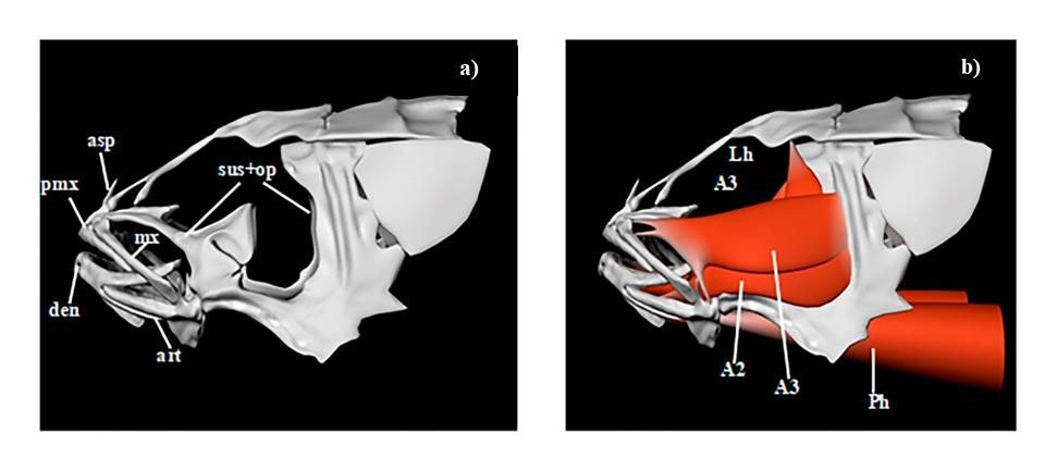
Fig. 3.
a) Three-dimensional solid model of the fish viscerocranium (lateral view);
b) Three dimensional osteomuscular model of the fish viscerocranium (lateral view). Designations of the main elements: Bones: art – anguloarticulare, den – dentale, mx – maxillare, op+sus – suspensorium + operculum complex, pmx – praemaxillare (asp – ascending process of the pmx). Musculature: A1, A2, A3 – portions of m. adductor mandibulare, Lh – m. levator hyomandibulare et arcus palatini, Ph – m. protractor hyoidei.
Assembly of section contours into a wireframe model was carried out in the Femap V 10 software [22] (Fig. 2 b). Using the geometric modeling command package (Parasolid, Standard), the wireframe models were transformed into solid counterparts (Fig. 2 c). The required elements of the viscerocranial bones and musculature were reconstructed based on visualization of fixed preparations of fish heads in the 3D modeling and animation software Autodesk Maya 2012 [23] (Fig. 3 a, b). The reliability of the geometric parameters of the resulting model was ensured by using morphometric data of the studied object. Generation of the finite element mesh in the solid model was implemented in the integrated environment of the design and modeling systems Patran [24].
Conditions for FEM testing. The main objective of this work was to determine and visualize the stress–strain state in an object of given shape, regardless of the properties of its material. The localization and direction of application of the test load were determined based on literature data characterizing the morphofunctional features of the fish jaw apparatus [25, 28]. Concentrated forces were applied at key nodes located at the attachment sites of the leading elements of the musculoligamentous apparatus of the head, as demonstrated on the osteomuscular virtual construction (Fig. 3 b). Additionally, loads simulating the pressure of prey on the tooth surface of the praemaxillare and dentale were modeled. The direction of the muscle force vectors was set according to previously obtained data from testing kinematic models of the fish jaw apparatus [29].
When assigning material properties, data on the
mechanical characteristics of bone tissue of the frontal bone of reptiles (E =
5600 Pa – elastic modulus, ν = 0.29 – Poisson's ratio) were used, which is due to
the lack of data on the mechanical characteristics of fish [19]. To identify zones
of maximum stress and potential deformations, a minimum load (10 N per node)
was applied to the model under study.
For the analysis of finite element models, a static
calculation was performed using the solver in the Nastran software [30], taking
into account the imposed boundary conditions and selected load variants.
Based on the analysis of the SSS of the obtained
objects, zones of potential adaptive deformations were identified for two
positions of the oral apparatus: 1) “preparation for prey capture” (mouth
opening and protrusion of the praemaxillare) and 2)
“prey capture” (mouth closing) (Figs. 4, 5). Visualization of the SSS
calculations was performed in the Patran environment,
in accordance with a color scale characterizing the distribution of stress
fields in the presented objects.
Position “Preparation for prey capture”: The mouth opening mechanism is ensured by the functioning of the opercular musculature, which acts through the l. mandibulo operculare on the dentale in the symphyseal region, causing its vertical downward displacement. The lower jaw block descends and moves the ventral ends of the praemaxillare and maxillare downward via the l. maxilla mandibulare anterius connecting to it. Through the A1 portion of the m. adductor mandibulae, a force is exerted on the cranial part of the maxillare, as a result of which it pushes forward the praemaxillare, which is movably articulated with the ethmoidal part of the neurocranium [25, 28]. The localization and distribution of the SSS during mouth opening for each of the jaw bones were as follows (Fig. 4):
• The praemaxillare, as a result of passive protrusion due to the action of the
maxillare, experiences compressive stress in the zone of their direct contact (the
articular process area);
• The maxillare experiences stress from axial force, which results from the action of the A1 portion of the m. adductor mandibulae attached near the articular head; the region of greatest stress is located in the compression zone (resistance to lateral bending), situated on the inner side of the bone;
• The dentale experiences local tensile stress in the attachment area of the l. maxilla-mandibulare, which lowers the lower jaw downward;
• The anguloarticulare experiences the greatest tensile stress at the base of the upper process, as a result of resistance to dorso-ventral bending of the lower process arising under the action of tensile forces from the dentale (contact between the anguloarticulare and the dentale).
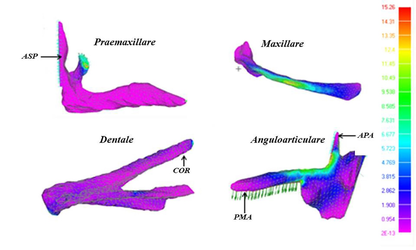
Fig. 4. Stress fields on the surface of the FEM of the jaw bones in the “mouth opening” position: Praemaxillare, asp – ascending process; Dentale, cor – coronoid process; Anguloarticulare, apa – upper process, pma – lower process. Note: green arrows indicate the localization and direction of the applied forces; light blue arrows indicate the boundary conditions.
Position “Prey capture”: Mouth closing is accomplished by the complex of the m. adductor mandibulae, which originates in the postorbital region of the viscerocranium; further, the muscle bundles attach in the area of the upper process of the anguloarticulare (A2) and the coronoid process of the dentale (A1, A2, A3, and Aw). The adductor complex, acting on these processes, moves the lower jaw upward. As the lower jaw rises, it pulls the ventral ends of the praemaxillare and maxillare via the l. maxilla mandibulare anterius, returning them to their previous position [20, 23]. The localization and distribution of the SSS during prey capture and mouth closing for each of the jaw bones were as follows (Fig. 5):
• The praemaxillare, under the pressure of the held prey, experiences compressive stress in the horizontal plane of the bone, as a result of its resistance to
lateral bending; the maximum compressive stresses are concentrated in the region of the upper process, which undergoes dorso-ventral bending;
• The maxillare experiences the same loads as in the previous variant, due to the action of similar forces;
• The dentale experiences maximum stresses in the region of the coronoid process, due to resistance to deformations of the “torsion” type, which result
from the combined application of several multidirectional forces: compression from the tooth surface (resistance of the prey), tension of the process from the m. adductor mandibulae complex, and compression arising from the resistance of the lower process of the anguloarticulare;
• The anguloarticulare experiences the greatest bilateral stress at the base of the upper process, as a result of its resistance to dorso-ventral
bending arising under the action of tensile forces from the A2 portion of the m. adductor mandibulae.
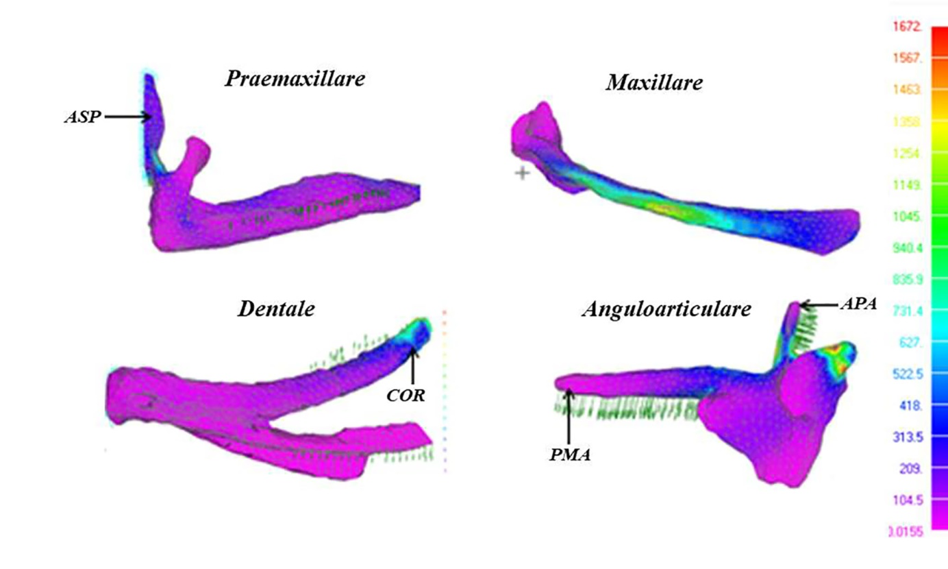
Fig. 5. Stress fields on the surface of the FEM of the jaw bones in the “mouth closing” position: Praemaxillare, asp – ascending process; Dentale, cor –
coronoid process; Anguloarticulare, apa – upper process, pma – lower process. Note: green arrows indicate the localization and direction of the applied forces;
lightblue arrows indicate the boundary conditions.
Functional analysis of structural transformations of
the feeding apparatus is inextricably linked to the issues of formation of the
trophic strategy of a particular species. Analysis of numerous literature data
shows that the leading trend in morphological variability within most fish
families is the elongation of the jaws and reduction of mouth protrusion, which
is associated with an increase in the size of food objects and a partial
transition to ichthyophagy [30-35]. This trend was previously noted for a group of closely related species of coastal
Cottoidei, demonstrating a consistent shift in trophic
adaptation from benthophagy to ichthyophagy (in the direction:
Paracottus knerii – Leocottus kesslerii – Batrachocottus baicalensis) [34].
Using graphical visualization of morphometric data of the jaw bones, the
interrelated variability of a number of morphogeometric parameters was revealed and demonstrated
(Fig. 6). It was established that the main trend in the transition from benthophagy to
ichthyophagy is the elongation of the jaws and a decrease
in their horizontal parameters, in particular, a decrease in the height of the
ascending process of the praemaxillare – AC (from
80.7 to 51.6 in % of bone length), a decrease in the angle between the plane of
the anguloarticulare and its upper process – ∠CDA (from 88° to 66°), and a decrease in the height of
the dentale – BC (from 43.4 to 35.4 in % of bone
length) (Fig. 6). It is known that bone remodeling occurs at sites of greatest
stress, which results from the action of the musculoligamentous apparatus [1]. The results of the present study are consistent with previously
obtained data. As shown above, the maximum stress fields tend to concentrate in
areas undergoing the greatest modification, in particular in the region of the
jaw processes, which serve as attachment points for muscles and ligaments.
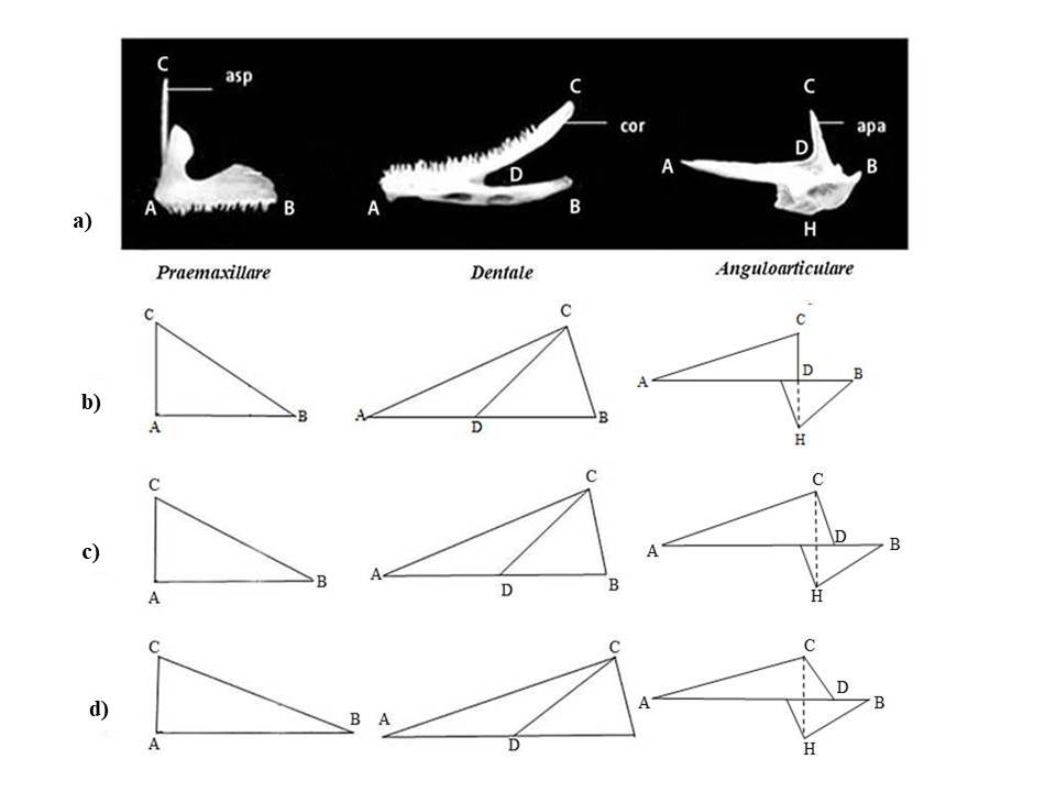
Fig. 6.Geometric projections of the jaw bones of Baikal sculpin fishes of the littoral group (Cottoidei): a) measurement scheme (in % of bone length); b) P. knerii; c) L.kesslerii; d) B.baicalensis [35];
Designations: Praemaxillare: asp – ascending process, (AB – bone length, AC – height of asp);
Dentale: cor – coronoid process (AB – bone length, BC – height, AC – position of the posterior end of the coronoid process, DC – length of the coronoid process);
Anguloarticulare: apa – upper process (AB – bone length, AD – position of the base of the upper process, CD – length of the upper process, CH – bone height, ∠CDA – angle formed by the upper process and the bone plane).
According to experimental
data, for effective prey capture, the upper jaw of fishes is protruded forward,
which occurs through the sliding of the ascending processes of the
praemaxillare along the ethmoidal part of the skull [26-34].
It is evident that the maximum force load during prey capture falls on the
ascending processes of the praemaxillare, analogous
to the “redistribution of the foot's supporting load to concentration in the
heel region.” Accordingly, with the elongation of the praemaxillare
and the “support” area in predators, the pressure on the ascending processes
increases, which leads to their gradual reduction (Figs. 4-6).
With jaw elongation in ichthyophages, greater effort is required for mouth opening/closing [1]. This process is accomplished by the A2 and AW portions of the m. adductor mandibulae complex, which attach to the inner side of the body of the anguloarticulare and its upper process [26, 27]. The increasing force load from the musculature leads to modification of the shape of the anguloarticulare. Thus, in the case of a “perpendicular” orientation of the process in benthophages, the forces of the m. adductor mandibulae A2 are unidirectional with the cross-section and act mainly on the longitudinal axis (Fig. 6). The successive deviation of the upper process toward the plane of the anguloarticulare during the transition to ichthyophagy leads to a redistribution of stress between its longitudinal and cross-sections due to their displacement relative to the direction of forces, and consequently reduces the risk of irreversible deformations under increased load (Fig. 5). The displacement of the upper process of the anguloarticulare into a horizontal position entails a decrease in the height of the dentale, which contacts it in the region of the coronoid process, experiencing a combined effect from the m. adductor mandibulae complex and the captured prey.
The development of methods for
virtual reconstruction of anatomical structures makes it possible to visualize
and reveal hidden patterns of transformation of the skeleton and its individual
elements, which are not always evident when using classical morphometry. Of
great interest in studying the adaptive variability of the jaw apparatus in
various taxa of vertebrates is the functional analysis and visualization of the
variability of the shape of skeletal structures and the prediction of their
behavior under the action of external forces. In the present work, to solve the
problem of the stress–strain state of the object, one of the modern
mathematical approaches – the finite element method – was applied. The
development and visualization of the FEM of the fish jaw apparatus enabled the
visualization and identification of the localization of areas with the greatest
stress under simulated loading conditions. Based on the obtained results, it
was established that structural changes occur at sites of maximum bone stress
concentration, with the jaw processes, which play a key role in the functioning
of the fish oral apparatus, being subject to the greatest deformation. It is
evident that the particular features of transformations of the jaw bones in
different fish species are a consequence of the uneven action of force loads,
which change during adaptation to feeding on different groups of organisms.
The results obtained at the
current stage of the research require further development, in particular, the
determination of the mechanical properties of fish bone material and its
integration with the features of shape variability of the object under load.
1. Wolff.J. Das Gesetz der Transfomation der Knochen. Berlin: Hirschwald. 1892. 235p.
2. Cowin S.C. Wolff’s law of trabecular architecture at remodeling equilitrium. J. Biomech. Engng. 1986. V. 108. P. 83-88.
3. Dumont E.R., Grosse I.R., Slater G.J. Requirements for comparing the performance of finite element models of b Biological structures. J Theor Biol. 2009. V. 256. P. 96–103. doi:10.1016/j.jtbi.2008.08.017.
4. O'Higgins P., Fitton L., Phillips R., Shi J.F., Liu J., Groening F., Cobb S.N., Fagan M.J. Virtual functional morphology: novel approaches to the study of craniofacial form and function. Evolutionary Biology. 2009. doi: 10.1007/s11692-012-9173-8.
5. Kupczik K. Virtual biomechanics: basic concepts and technical aspects of finite element analysis in vertebrate morphology. Journal of Anthropological Sciences. 2008. V. 86. P. 193–198.
6. Ross CF. Finite element analysis in vertebrate biomechanics. Anat Rec A Discov Mol Cell Evol Biol. 2005 Apr;283(2):253-8. doi: 10.1002/ar.a.20177. PMID: 15754323.
7. Richmond B. G., Wright W., Grosse I., Dechow P. C., Ross C., Spencer M., and Strait D. Finite element analysis in functional morphology. Anat. Rec. 2005. V. 283. P. 259–274. doi: 10.1002/ar.a20169.
8. de Simao-Oliveira D, Dos Santos T, Pinheiro FL, Pretto FA. Assessing the adductor musculature and jaw mechanics of Proterochampsa nodosa (Archosauriformes: Proterochampsidae) through finite element analysis. Anat Rec (Hoboken). 2024 Apr;307(4):1300-1314. doi: 10.1002/ar.25380.
9. Melki LB, Barbosa FHS, Alves-Silva L, Bergqvist LP. Variations in dietary patterns of living sloths revealed by finite element analysis of jaws. J Anat. 2024 Oct;245(4):583-592. doi: 10.1111/joa.14099.
10. Panagiotopoulou O, Robinson D, Iriarte-Diaz J, Ackland D, Taylor AB, Ross CF. Dynamic finite element modeling of the macaque mandible during a complete mastication gape cycle. Philos Trans R Soc Lond B Biol Sci. 2023 Dec 4;378(1891):20220549. doi: 10.1098/rstb.2022.0549.
11. Wilken AT, Schultz JA, Luo ZX, Ross CF. A new biomechanical model of the mammal jaw based on load path analysis. J Exp Biol. 2024 Sep 15;227(18):jeb247030. doi: 10.1242/jeb.247030.
12. Watson PJ, Sharp AC, Choudhary T, Fagan MJ, Dutel H, Evans SE, Groning F. Computational biomechanical modeling of the rabbit cranium during mastication. Sci Rep. 2021 Jun 23;11(1):13196. doi:10.1038/s41598-021-92558-5.
13. Penrose F, Cox P, Kemp G, Jeffery N. Functional morphology of the jaw adductor muscles in the Canidae. Anat Rec (Hoboken). 2020 Nov;303(11):2878-2903. doi: 10.1002/ar.24391.
14. Nieto-Miranda JJ, Aguilar-Medrano R, Hernandez-Camacho CJ, Peredo CM, Cruz-Escalona VH. Mechanical properties of the California sea lion (Zalophus californianus) and northern elephant seal (Mirounga angustirostris) lower jaws explain trophic plasticity. Anat Rec (Hoboken). 2023 Oct;306(10):2597-2609. doi: 10.1002/ar.25180.
15. Peterson T, Muller GB (2018) Developmental finite element analysis of cichlid pharyngeal jaws: Quantifying the generation of a key innovation. PLoS ONE 13(1): e0189985. https://doi.org/10.1371/journal.pone.0189985
16. Brunt LH, Roddy KA, Rayfield EJ, Hammond CL. Building Finite Element Models to Investigate Zebrafish Jaw Biomechanics. J Vis Exp. 2016 Dec 3;(118):54811. doi: 10.3791/54811.
17. Tolmacheva Yu.P., Dolid E.A., Petukhov S.Yu., Pashkov V.P., Pykhalov A.A. 3D modeling and animation of the visceral skeleton of fish: testing the system of four-link mechanisms. Mathematical biology and bioinformatics. 2013. Vol. 8. No. 2. P. 513–519. doi: 10.17537/2013.8.513.
18. Tolmacheva, Yu.P., Shmatkova A.V., Zaides S.A. Application of the finite element method for visualization of the stress-strain state of fish bones// Scientific visualization. – 2019. – T. 11, No. 2. – P. 114-125. – DOI 10.26583/sv.11.2.
19. iCATVision 1.8 Developer by ConeBeam Imaging LLC. // Software for visualization of CT scans of the maxillofacial area URL :https://icatvision.software.informer.com/1.8/ (date of access 05/02/2018)
20. XnView 1.98.2 Developer by XnSoft. // Software for viewing and editing images URL : https://www.xnview.com/en/ (date of access 15/02/2018)
21. Femap V 10 Developed by Siemens Product Lifecycle Management Software Inc. //System of finite element modeling and postprocessing application URL: http://www.femcomp.com/ (date of access 15/02/2018)
22. Autodesk Maya 2012 Developed by Autodesk, Inc // Software for 3D modeling and animation URL: http://www. autodesk.com/ (date of access 10/03/2018)
23. Patran Developed by MSC Software // System for integrating automated design, modeling, analysis, and evaluation of calculation results. URL: http://www.patran.net/ (date of access 15/02/2018)
24. Dobben W.N. Uber der Kiefermechanismus der Knochenfishe. Archive neerland. Zoolog. 1935. V. 50. P. 1–72.
25. Yabe M. Comparative osteology and miology of the superfamily Cottoidea (Pisces: Scorpaeniformes), and its phylogenetic classification. Reprinted from Memoirs of the Faculty of Fisheries, Hokkaido University. 1985. V. 32. P. 130.
26. Westneat M.W. Feeding mechanics of teleost fishes (Labridae: Perciformes): A test of four-bar linkage models. J. Morph. 1990. V. 205. P. 269–295. doi: 10.1002/jmor1052050304.
27. Yu. P. Tolmacheva, A. V. Shmatkova, D. T. Wu, V. V. Kuzlyakina. Functioning of the jaw apparatus of fish: modeling and kinematic analysis of the crank-rocker mechanism // Mathematical biology and bioinformatics. - 2018. - Vol. 13, No. 2. - P. 551-562. DOI 10.17537/2018.13.551.
28. Gregory W.K. Fish skulls. A study of the evolution of natural mechanism. Trans. Amer. Phil. Soc. 1933. V. 23. P. 75-481.
29. Nastran Developed by MSC Software // Software for finite element analysis (FEA) URL: http://www.mscsoftware.ru/products/msc-nastran/ (date of access 17/04/2018)
30. Gosline W.A. Comments on the classification of the percoid fishes. Pacific. Sci. 1966. V. 4. P. 409-418.
31. O’Kamura O. Stadies of the Macrouroid fishes of Japan. Morfology, ecology and phylogeny. Rept. U. S. Mar. Biol. Stat. 1970. 32 p.
32. Voskoboynikova O.S. Evolutionary transformations of the visceral skeleton and questions of phylogeny of nototheniidae fishes. Tr. Zool. Institute of the USSR Academy of Sciences. 1986. T. 153. pp. 46-66.
33. Tolmacheva, Yu. P. Structure of the oral and pharyngeal apparatus of three species of Baikal Cottoidei in connection with their nutrition / Yu. P. Tolmacheva // Questions of ichthyology. - 2010. - V. 50, No. 1. - P. 69-76.
34. Tolmacheva, Yu. P., Bun M. M. Structure features and structural transformations of the maxillary bones in the Baikal cottoid fish // Limnology and Freshwater Biology. – 2019. – No. 5. – P. 308-311. – DOI 10.31951/2658-3518-2019-A-5-308.