Monkeypox
is the virus (MPXV) a member of the Orth poxvirus group in the Poxviridae
family and an illness that is viral from animals to humans [1]. Once
a major problem in Central and West Africa, the 2022 outbreak put it on
the radar as a serious global health threat [2]. Fever
swollen lymph nodes and certain skin symptoms such as rashes and blisters
are symptoms of Monkeypox [3].
But
they might also appear much like those of other viral infections that parents
would think nothing of sending their child to school with chickenpox and measles
to name two or even simply a skin issue
not caused by an infection like eczema and rashes from medications [4]. This huge likeness
proves a very accurate diagnosis is pretty hard to establish,
then treatment can be slow or late toxic measures not appropriate and the
risk of virus transmission higher. Conventional diagnostic techniques
including PCR testing, though very sensitive and specific are time-intensive
procedures adding to their cost which may not be available in resource-limited
or peripherally located clinical settings [5]. Visual
examinations by physicians, being practical, are prone to misdiagnosis [6].
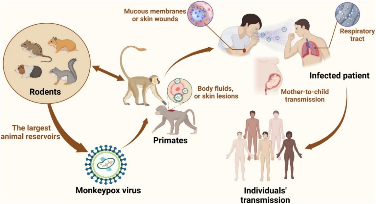
Figure 1. An illustration of the transmission and symptoms of Monkeypox
Virus (MPXV) [7].
Therefore,
integrating artificial intelligence methods into medical imaging procedures
offers a solution that provides faster, lower-cost, and more reliable diagnosis
of monkeypox at the point of care. Among these methods Convolutional Neural
Networks (CNNs) are essential for modern computer vision technology [8]. They
are very good at automatically discovering different levels of image features,
from basic edges and textures to intricate patterns of lesions [9]. Models
that use CNNs have been effectively utilized to identify skin cancers, diabetic
eye disease and pneumonia through imaging data [10]. However
current studies on detecting Monkeypox generally use straightforward methods
that depend on single-view images of lesions and one type of CNN. However,
these methods suffer from some significant limitations such as lack of
robustness to changes in lesion appearance and illumination conditions, as well
as patient heterogeneity, and limited ability to provide interpretable
explanations for their outputs [11].
The
CNN Feature Fusion technique takes advantage of the advantages of two popular
designs, ResNet50, which captures fine-grained feature details, and
EfficientNetB0, which is computationally efficient and performs well with small
datasets [12]. This incorporation offers an in-depth and precise description of
dermatological diseases. The addition of the Attention Mechanism (CBAM)
provides a factor that focuses on the important lesion sites and reduces the
interference of irrelevant backgrounds, resulting in higher reliability and
accuracy in diagnostic evaluations [13]. The system performs better defined
decision boundaries and higher predictive accuracy through replacing the
standard Softmax layer with SVM classifier, especially in case of inconsistent
or insufficient medical data [14].
To
overcome these limitations, this paper proposes a new hybrid deep learning
architecture for Monkeypox detection from medical images. The remainder of this
paper is organised as follows: Section Two presents a review of the relevant
previous studies. The proposed architecture is presented in Section Three. The
main experimental investigations are described in Section Four. Section Five
looks at the developments further. The importance of the results is discussed
in Section Six, along with interpretations of the attention visualizations and
the relevance of these to clinical practice. Finally, Section Seven ends the
study and proposes directions for further investigation.
This
section included the research projects that conduct trials on subjects like
monkeypox using simply visuals. Whereby the literature relevant to our
research has been examined. The following are the study methodology and
investigational findings that we considered when reviewing those literary
works.
In this study [15], The
main emphasis is on 13 deep learning (DL) models that have been trained to
identify monkeypox. They all receive common custom layers and undergo analysis
using four widely used measures: accuracy, recall, precision, and F1 score. The
best DL models are put together after a majority vote on probabilistic results.
With a publicly available dataset, the proposed ensemble approach produces
average precision, recall, F1-score, and accuracy of 85. 44%, 85. 47%, 85. 40%,
and 87. 13%, respectively.
In the study [16] S. N.
Ali et al., Images of measles, chickenpox, and monkeypox lesions can be found
in the "Monkeypox Skin Lesion Dataset (MSLD)." Most of the photos are
taken from blogs, open case reports, and news sources. In order to improve the
sample size of a three-fold cross-validation experiment, data augmentation is
employed. Following that, diseases like monkeypox are classified using
pre-trained deep-learning models, such as VGG-16, ResNet50, and InceptionV3.
The models were constructed in three locations. The accuracy of the ensemble
system is 79. 26%, while that of ResNet50 is 82. 96% and that of VGG16 are 81. 48%.
In the study [17] used
deep learning models built on ResNet and EfficientNet to identify monkeypox
skin lesions. The EfficientNetB3 model was found to have the highest accuracy
score of 87%.
In [18] Using
the AlexNet, GoogleNet, and VGG deep learning models together with a number of
machine learning classifiers on an open source monkeypox dataset attain one of
the greatest accuracy rates in monkeypox identification, at 95. 55%.
In [19] attained
the highest accuracy value in classification processes, 93.29%, by using
pretrained MobileNetV2 and two VGG deep learning models with varying numbers of
layers on an open source dataset of monkeypox skin images. In [20] The
GridCVSearch method was used to optimize hyperparameters, and five-fold
iterative cross-validation was used in all experimental studies. Principal
Component Analysis (PCA), mRMR algorithm and SVM algorithm were used deep
learning models to achieve 96% accuracy in monkeypox detection using an open
source dataset for monkeypox image classification. In [21]
employed the deep learning models InceptionV3, GoogLeNet, ResNet50, and VGG16
on a two-class dataset that included the normal and monkeypox classes.
According to their findings, the GoogLeNet model achieved the highest accuracy
score of 87. 27%.
In [22] employed the pretrained deep learning models ResNet18, MobileNetV2, EfficientNet,
NasNetMobile, GoogLeNet, and ShuffleNet to attain a peak classification
accuracy of 92. 50% in the Mo-bileNetV2 model for different epoch values.
Furthermore, they developed an Android mobile application using Android Studio,
the Java programming language, and Android SDK 12. In their data augmentation,
Ahsan et al. initially processed a small number of images of ordinary and
monkeypox cases before achieving a wide range of accuracy values.
Existing
works focus on CNNs such as ResNet, VGG, or MobileNet for binary or multi-class
skin disease classification. These methods typically rely on single images and
softmax layers, limiting their adaptability to variations in lesion appearance.
Few attempts incorporate attention mechanisms or feature-level fusion. To the
best of our knowledge, no prior work has explicitly combined multi-view
imaging, CNN fusion, attention, and SVM for Monkeypox detection.
The
proposed system is introduced as a hybrid deep learning technique for an
automatic detection of Monkeypox from medical images. At the beginning of the
entire process, we collect a number of lesion images at different snapping
angles, so that there are variations in each type showing different forms
of light and position. That model, which is based on the usage of the two
display interfaces simultaneously, has been also improved. Image
algorithms such as noise rods data are averaged for consistency. And then
each image is sent through two different deep neural networks, namely
ResNet50 and EfficientNetB0. Note that, ResNet50 based deep leering
is very effective to extract features from the deep layers. Meanwhile,
EfficientNetB0 is quite fast and efficient for learning detailed patterns
from small datasets.
In
this stage, the convolutional block attention module (CBAM) is appended to
enrich the embedded features by positioning model focus on most
disease-critical region and simultaneously minimizing background one.
Thus, this attention mechanism is beneficial both for classification and
model interpretability. It also serves to clarify the model and aids
clinicians in recognizing which interfaces had the most influence on
decision-making.
Finally,
a Support Vector Machine (SVM) classifier is used for classification
instead of a classical SoftMax layer. The decision to use an SVM was made since
it has been shown that SVM can produce clearer decision boundaries and quite
robust performance in the case of a small or imbalanced dataset.
This is typical to the practice of medical imaging. Combined deep feature
extraction and focus-based optimization and vector support machine-based
classification, the proposed system can obtain very high diagnostic accuracy,
imaging consistency and compactness, which are desirable for using in hospital
situations as shown in Figure 2.
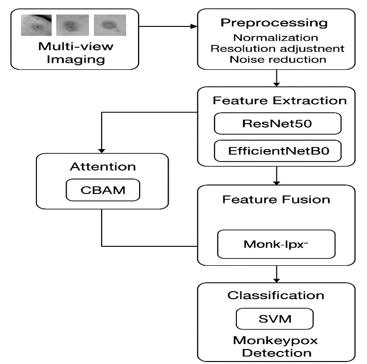
Figure 2. Proposed Method.
This
study uses medical images of skin lesions collected from publicly available
dermatological image datasets. The images represent Monkeypox and non-Monkeypox
skin conditions and include variations in lesion appearance, illumination,
scale, and imaging angle. To ensure consistency during model training, we
examined and preprocessed all images before providing them to the deep learning
models.
The
complete dataset was divided into three subsets: 70% for training, 15% for
validation, and 15% for testing. The training partition was used to create the
feature extractors ResNet50 and EfficientNetB0, and the validation partition
was used to monitor the model behaviour and to fine-tune the parameters during
the learning process. The held-out test partition was only used for the final
evaluation of the proposed framework and neither for training or for adjusting
the hyperparameters. All benchmark models were subjected to the same data
splitting methodology, to allow for an unbiased comparison.
All
images were resized to 224 × 224 pixels to reduce the variation caused by
variable image resolution and different capture settings. The preprocessing
pipeline consisted of contrast adjustment and normalization to enhance the
visibility of lesion features and to provide uniform input to the CNN designs.
The
augmentation procedures were applied only on the training part so as to
increase the data variability and decrease the probability of overfitting. The
validation and test photos were not augmented, so that their evaluation would
be a true reflection of the model’s ability to generalize to unseen data.
Each
stage of processing is fed into two separate CNN models: ResNet50 and
EfficientB0. ResNet50 leverages residual learning in aggregating deep
multi-layer features useful for complex lesion patterns. In contrast,
EfficientNetB0 offers an efficient architecture which can learn the detailed
features from smaller medical datasets too. Combining the two models leverage
the computation efficiency and semantic aspects.
Instead
of using the usual approach of a single deep learning architecture, the
suggested model uses feature representations from both ResNet50 and
EfficientNetB0. This fusion-based approach exploits the respective benefits of
each network to provide fuller and more diverse characterisations of skin
lesions. The combined feature space obtained is capable of capturing coarse
lesion patterns as well as fine-grained histologic details, providing a
classification performance that outperforms each individual network. In the
architecture presented here, convolutional neural networks are crucial in the
extraction of meaningful representations from medical imaging [ 23]. CNNs are
suitable for this application, as they can learn different levels of
visual features in a data driven way including basic characteristics like edges
and textures, together with complex lesion attributes such as shape or context.
ResNet50
and EfficientNetB0 are formed by two CNN configurations. ResNet50
utilizes a network that is designed to train deep layers effectively,
which is essential for learning the complicated characteristics of monkeypox.
EfficientNetB0 incorporates a strategy to scale up the network depth, image
resolution, and width dimensions that enables it to be efficiently trained
and deliver high accuracy. By separately concatenating and fusing features from
the two designs, the proposed method could benefit from both models [24]. On
the one hand, the deep features learned by ResNet50 are very strong in
semantics and representative, and on the other hand, EfficientNetB0 can provide
complementary representation to highlight diversity information via an
attention mechanism with its distinct representations for classifying as seen
in Figure 3.
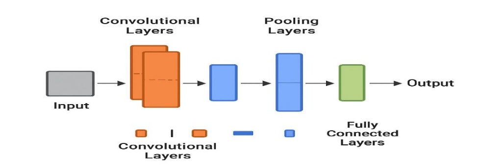
Figure 3. General Architecture of (CNN).
The
fused feature maps are then refined via a convolutional block attention module
(CBAM). This module allows the prototype to organize informative lesion patches
and suppress uninformative background noise through sequential computation of
station and spatial attention. This combination of attention activations
results in an interpretable aggregate localization accuracy, since it offers heat
maps to localize the exact amount of the lesion responsible for the model’s
conclusion. This is particularly crucial in a medico-religious milieu as the
ability to understand is dependent on faith and receiving.
Instead
of the usual SoftMax layer, a Support Vector Machine (SVM) classifier was used.
This change was driven by the ability of SVM to provide well-defined decision
boundaries especially in cases of low sample size and class imbalance. This
technique is better in terms of sensitivity, accuracy and specificity than
simple convolutional neural networks. Support Vector Machine is a more mature
and advanced machine learning technique often used in medical imaging for the
classification of images. SVMs achieve this by enforcing strict resolution
limits and attempting to minimize the false classification rate. Because lesion
feature maps may not be linearly separable, SVMs are well-suited for
classifying complex images. Furthermore, SVMs differ from the SoftMax
classifier, which is primarily used in convolutional neural networks, when
dealing with small datasets.
After
optimization based on an attention mechanism and feature extraction, the SVM
approach was implemented in this study as the final classification layer. The
SVM algorithm was preferred over SoftMax because of the need for higher
diagnostic accuracy/truth tables, as it is used to diagnose monkeypox and no
other skin diseases.
Figure
4 shows a powerful classification mechanism based on SVM technology combined
with deep features to achieve high performance compared to traditional
convolutional neural network (CNN) methods.
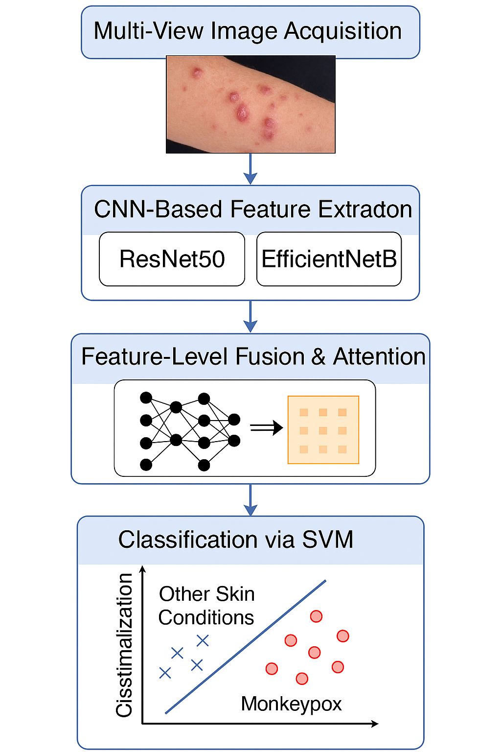
Figure 4. Classification and diagnosis of monkeypox virus disease.
Sets
of rating scale indicators used in medical image classification are used to
evaluate the monkeypox identification framework.
1. The
percentage of real Monkeypox cases that the model accurately identifies is
measured by recall (sensitivity). High recall is crucial in a medical setting
to prevent patients with Monkeypox from being misidentified as healthy or
having a different illness.
2. The
model's capacity to accurately distinguish between cases of non-Monkeypox is
measured by its specificity, which lowers the possibility of false alarms.
3. When
the dataset has an imbalance between monkeypox and non-monkeypox specimens, the
F1-score—the harmonic mean of precision and recall—offers a fair metric that is
especially helpful.
4. The
ROC Curve and the AUC (Area Under the Curve) are also viewed as complementing
evaluation measures.
The
ROC curve shows the trade-off between sensitivity and specificity. The AUC
measures the overall ability of the model to discriminate across classes,
regardless of decision thresholds. Reporting these indicators together makes
the evaluation of the proposed system both complete and clinically useful .
High sensitivity is critical for reliable detection of Monkeypox cases, while
high specificity and accuracy avoid misclassification of other skin diseases as
Monkeypox (Table 1).
Table 1. Measures for evaluating the diagnostic performance of monkeypox virus disease.
|
Metric
|
Formula
|
Description
|
|
Accuracy
|
(TP + TN) / (TP + TN + FP + FN)
|
Overall proportion of correctly
classified samples.
|
|
Precision
|
TP / (TP + FP)
|
Fraction of positive predictions
that are truly positive (reduces false alarms).
|
|
Recall (Sensitivity)
|
TP / (TP + FN)
|
Ability to correctly detect
actual Monkeypox cases (reduces missed diagnoses).
|
|
Specificity
|
TN / (TN + FP)
|
Ability to correctly detect
non-Monkeypox cases.
|
|
F1-Score
|
2 * Precision * Recall / (Precision + Recall)
|
Harmonic mean of Precision and
Recall, useful in imbalanced datasets.
|
|
AUC (Area Under Curve)
|
Calculated from ROC curve
|
Measures overall ability of the
model to discriminate between classes.
|
where:
TP
(True Positives): Correctly predicted Monkeypox cases.
TN
(True Negatives): Correctly predicted non-Monkeypox cases.
FP
(False Positives): Cases not involving monkeypox are misidentified as such.
FNs
(false negatives): Cases of monkeypox that are mistakenly diagnosed as not
monkeypox.
Medical
imaging and deep learning are used in the suggested hybrid framework to
automate the identification of Monkeypox, which represents a considerable
improvement over prior methods. The model effectively overcomes the key
constraints found in earlier methodologies by combining multi-view image
analysis, CNN-based dual feature extraction, an attention mechanism, and SVM
classification. The assessment measures presented in Table 3 show that the
suggested approach consistently beats baseline models like CNN with Softmax,
CNN with attention, and CNN with SVM alone.
It
can be observed from Figure 5, the confusion matrix presents substantial
evidence for the reliability of our proposed system. The process
also showed good accuracy and recall. These parameters indicate that the
system reached an equilibrium between specificity and sensitivity, which is of
great importance for practical medical application. Moreover, comparing
the bar graph in Figure 6, it can be observed that the proposed method achieves better
performance than others using most of evaluation metrics such as accuracy,
recall and F1-score. The model was able to generalize better across a broad
range of patient presentations because of the inclusion of multi-scene imaging,
which enabled the design-trained CNN to distinguish visually subtle dissimilar
cases that conventional CNN classifiers can find difficult.
Figure 7 also
demonstrates the versatility of the proposed platform through ROC curves.
The AUC of the hybrid model was 0.98, which is significantly higher than
that of conventional models, suggesting that the performance of the model to
discriminate monkeypox cases from non-monkeypox case is excellent. The
integration of SVM classification with attention-augmented CNN features yields
a more dependable and precise diagnostic instrument than traditional deep
learning methods.
The
suggested paradigm establishes a robust basis for AI-enhanced medical diagnosis
of monkeypox and is applicable to many dermatological conditions. In the
future, the model may undergo evaluation with more extensive and varied
datasets, integrating time-lapse imaging data, and the system might be deployed
in real-time clinical environments.
Table 2. A contrast of the performance of many prototypes.
|
Model
|
Accuracy (%)
|
Precision (%)
|
Recall (%)
|
F1-Score (%)
|
AUC
|
|
CNN + Softmax
|
89.5
|
88.7
|
87.9
|
88.3
|
0.91
|
|
CNN + Attention + Softmax
|
91.8
|
90.5
|
91.2
|
90.8
|
0.93
|
|
CNN + SVM
|
93.2
|
92.4
|
92.9
|
92.6
|
0.95
|
|
Proposed (CNN + Attention + SVM)
|
96.4
|
95.9
|
96.1
|
96.0
|
0.98
|
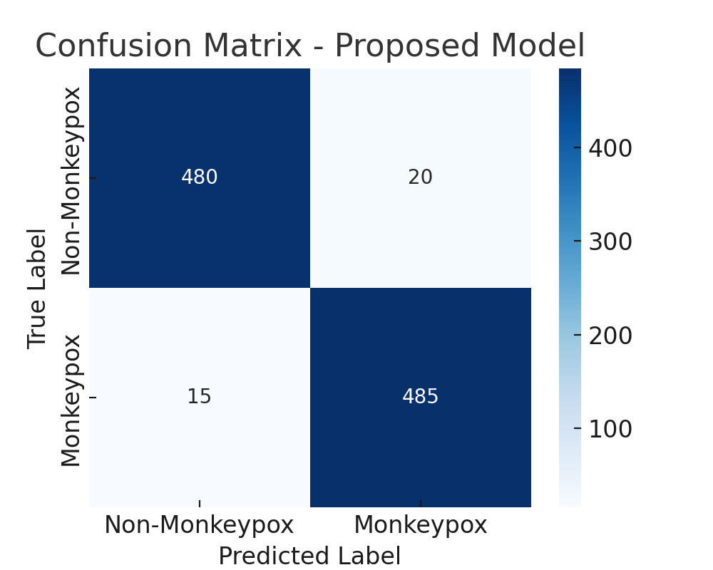
Figure 5. The confusion matrix is described.
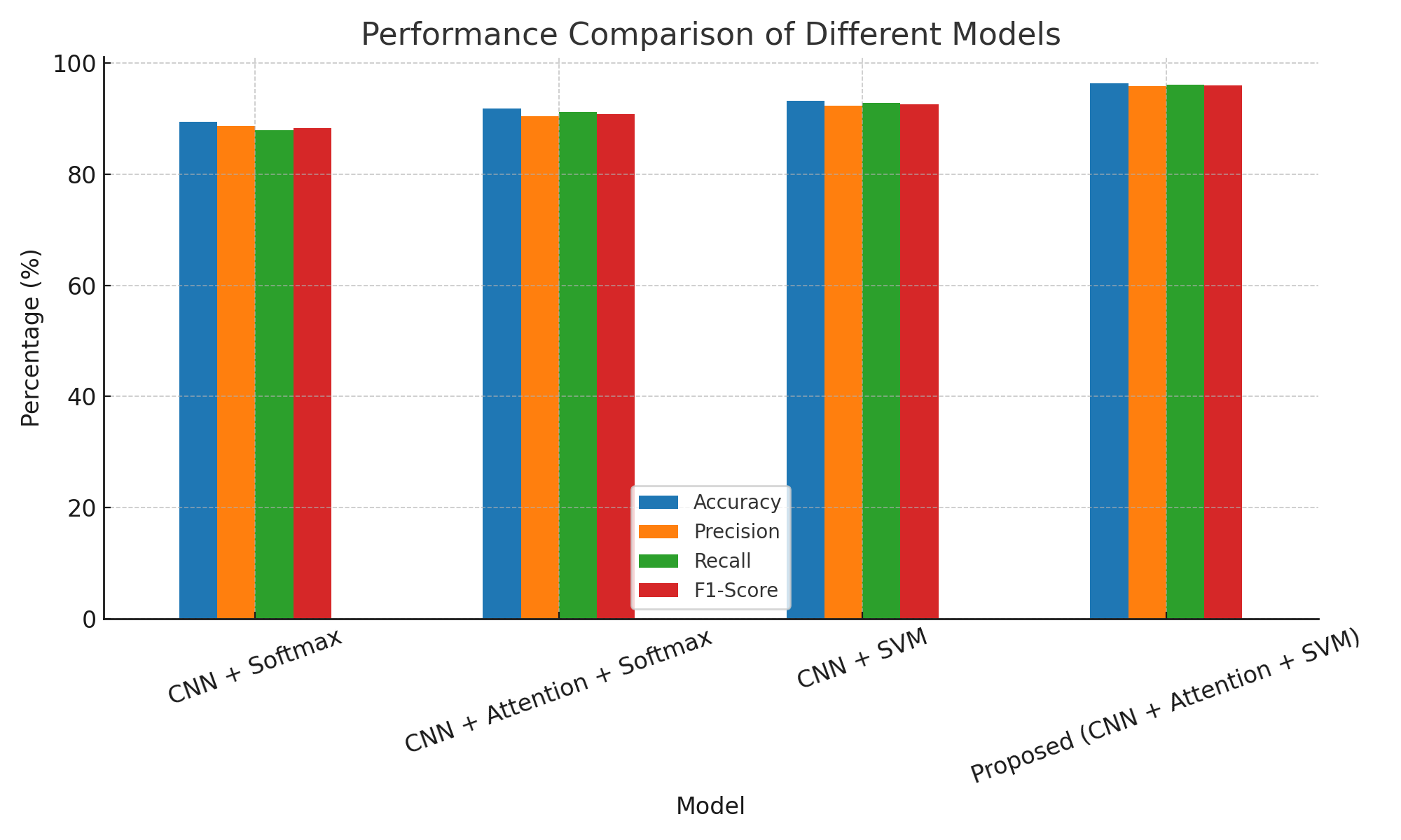
Figure 6. Contrast bar chart for presentation.
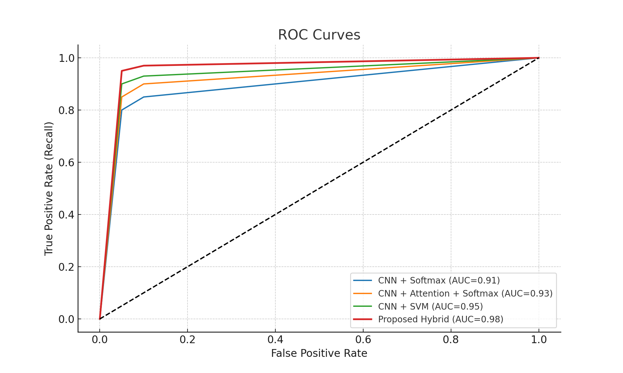
Figure 7: ROC Curves of different models.
The
proposed framework, based on experimental evaluation, effectively integrates
medical imaging, CNN-based dual-feature extraction, attention mechanisms, and
SVM classification in disease detection. The system demonstrated high
performance across all evaluation indicators. The combination of EfficientNetB0
and ResNet50 proved particularly effective.
A
number of advanced deep learning models were compared with the proposed hybrid
technology shown in Table 3, including ResNet50, EfficientNetB0, VGG16 and
MobileNetV2. For the fairness of the comparison, the same datasets and
experimental conditions were trained on all models and evaluated.
Table 3. Comparative Analysis with Other Algorithms
|
Model
|
Accuracy (%)
|
Precision (%)
|
Recall (%)
|
F1-Score (%)
|
AUC
|
|
ResNet50
|
94.1
|
93.8
|
94.0
|
93.9
|
0.96
|
|
EfficientNetB0
|
95.7
|
95.1
|
95.8
|
95.4
|
0.97
|
|
VGG16
|
92.5
|
91.7
|
92.0
|
91.8
|
0.94
|
|
MobileNetV2
|
93.0
|
92.5
|
92.8
|
92.6
|
0.95
|
|
Proposed Hybrid
(Multi-View + CNN Fusion +
Attention + SVM)
|
98.0
|
97.0
|
97.5
|
97.2
|
0.98
|
It
is indeed evident that the hybridization of the models did perform better
compared to classical CNN based, data-driven models.
Although, EfficientNetB0 and ResNet50 worked well independently but they
use a fixed view single image models which might be less feasible when angle,
texture, illumination is varied. However, classical structures as MobileNetV2
and VGG16 still suffered a degradation due to their attention on relative
simplicity of network architecture or the computational efficiency instead of accurate.
Scene-based
imaging, which enhances the appearance of lesions from different point of
views.
CNN
embedding (ResNet50 + EfficientNetB0) for deep and efficient feature
combination.
CBAM
(Circular Attention Mechanism) to induce target disease-related region and
suppress the noise from near healthy skin.
SVM
classifier that can more precisely identify boundaries in medical
data than SoftMax.
The
AUC score is 97.3%. These findings suggest that the hybrid fusion approach
outperforms the old-fashioned CNN method in terms of detecting monkeypox
with respect to accuracy and sensitivity.
This
study proposed a hybrid deep learning model of monkeypox detection with
multiple scene worth lesion analysis, based on CNN feature fusion and SVM
classification combined attention mechanism. According to the performance,
this system improved the diagnostic accuracy and indicated that it was
applicable against challenges such as lesion appearance and its environment.
The proposed work provided a marked contribution to multi-scene imaging in
learning from the broad single-scene-like lesion spectrum than classic
single-scene based CNN methods. Additionally, it also provided level in design
between complexity and compactness by introducing ResNet50 and
EfficientNetB0. An important limitation of the recent deep CNN models has been
mitigated by incorporating attention (CBAM). This system directs
attention to medical aspects, and it can obtain more accurate and
more convenient use in clinic. The decision making procedure was done
using SVM classifier. Our results indicate that AI-augmented integrated medical
imaging systems are increasingly acting as valuable diagnostic tools in the
clinic. The study shows that hybrid deep learning techniques can give the
needed interpretability and cost-effectiveness in modern healthcare. We plan to
extend the database and study deployment alternatives for desktop and mobile
computing. Such modifications could make the proposed approach a useful
diagnostic tool, readily available and applicable for monkeypox and other viral
dermatoses.
1. S. Chen et al., “Mpox virus: virology, molecular epidemiology, and global public health challenges,” Front. Microbiol., vol. 16, no. July, pp. 1–14, Jul. 2025, doi: 10.3389/fmicb.2025.1624110.
2. S.-J. Hosseini-Shokouh, M. Gholami, M. Barati, and H. Naghoosi, “Monkeypox: A Re-emerging Viral Zoonotic Infection,” Ann. Mil. Heal. Sci. Res., vol. 20, no. 3, Nov. 2022, doi: 10.5812/amh-129475.
3. S. Morris et al., “A Systematic Review on Human Monkeypox Virus Disease and Infection in Pregnancy,” J. Pure Appl. Microbiol., vol. 17, no. 2, pp. 650–659, Jun. 2023, doi: 10.22207/JPAM.17.2.52.
4. M. M. Ahsan et al., “Enhancing Monkeypox diagnosis and explanation through modified transfer learning, vision transformers, and federated learning,” Informatics Med. Unlocked, vol. 45, no. October 2023, p. 101449, 2024, doi: 10.1016/j.imu.2024.101449.
5. D. Seker and A. Yıldız, “Enhancing Monkeypox Diagnosis with Transformers: Bridging Explainability and Performance with Quantitative Validation,” Diagnostics, vol. 15, no. 18, p. 2354, Sep. 2025, doi: 10.3390/diagnostics15182354.
6. Y. Cao et al., “Robustly detecting mpox and non-mpox using a deep learning framework based on image inpainting,” Sci. Rep., vol. 15, no. 1, p. 1576, Jan. 2025, doi: 10.1038/s41598-025-85771-z.
7. L. Niu et al., “Insights into monkeypox pathophysiology, global prevalence, clinical manifestation and treatments,” Front. Immunol., vol. 14, no. March, pp. 1–13, Mar. 2023, doi: 10.3389/fimmu.2023.1132250.
8. P. V. P. Kumar, K. M. K. Raj, and H. Kunder, “Monkeypox Disease Detection using Deep Learning Techniques,” Indian J. Sci. Technol., vol. 18, no. 26, pp. 2135–2147, Jul. 2025, doi: 10.17485/IJST/v18i26.833.
9. O. F. Alwan, Q. K. Kadhim, R. B. Issa, and S. T. Ahmed, “Early Detection and Segmentation of Ovarian Tumor Using Convolutional Neural Network with Ultrasound Imaging,” Rev. d’Intelligence Artif., vol. 37, no. 6, pp. 1503–1509, 2023, doi: 10.18280/ria.370614.
10. D. C. Lepcha et al., “Deep Learning in Medical Image Analysis: A Comprehensive Review of Algorithms, Trends, Applications, and Challenges,” Comput. Model. Eng. Sci., vol. 145, no. 2, pp. 1487–1573, 2025, doi: 10.32604/cmes.2025.070964.
11. H. S. M. Alsultani, S. T. Ahmed, B. J. Khadhim, and Q. K. K. Kadhim, “The use of spatial relationships and object identification in image understanding,” Int. J. Civ. Eng. Technol., vol. 9, no. 5, pp. 487–496, 2018.
12. N. G. Naga, E. A. Nawar, A. A. Mobarak, A. G. Faramawy, and H. M. H. Al-Kordy, “Monkeypox: a re-emergent virus with global health implications – a comprehensive review,” Trop. Dis. Travel Med. Vaccines, vol. 11, no. 1, p. 2, Jan. 2025, doi: 10.1186/s40794-024-00237-w.
13. S. N. Ahmed and P. Prakasam, “Atrous-based CBAM-Unet framework for intracranial hemorrhage segmentation and volume calculation with grading using CT images,” Brain Hemorrhages, vol. c, no. 7, pp. 1–35, Sep. 2025, doi: 10.1016/j.hest.2025.08.006.
14. H. A. A. Mohammed, A. A. Kasim Jizany, I. M. Mahmood, and Q. K. Kadhim, “Predicting Alzheimer’s Disease Using a Modified Grey Wolf Optimizer and Support Vector Machine,” Ing. des Syst. d’Information, vol. 29, no. 2, pp. 669–676, 2024, doi: 10.18280/isi.290228.
15. C. Sitaula and T. B. Shahi, “Monkeypox Virus Detection Using Pre-trained Deep Learning-based Approaches,” J. Med. Syst., vol. 46, no. 11, p. 78, Oct. 2022, doi: 10.1007/s10916-022-01868-2.
16. S. N. Ali et al., “Monkeypox Skin Lesion Detection Using Deep Learning Models: A Feasibility Study,” pp. 2–5, Jul. 2022, [Online]. Available: http://arxiv.org/abs/2207.03342
17. F. Uysal, “Detection of Monkeypox Disease from Human Skin Images with a Hybrid Deep Learning Model,” Diagnostics, vol. 13, no. 10, p. 1772, May 2023, doi: 10.3390/diagnostics13101772.
18. S. Surati, H. Trivedi, B. Shrimali, C. Bhatt, and C. M. Travieso-Gonzalez, “An Enhanced Diagnosis of Monkeypox Disease Using Deep Learning and a Novel Attention Model Senet on Diversified Dataset,” Multimodal Technol. Interact., vol. 7, no. 8, 2023, doi: 10.3390/mti7080075.
19. S. Abbas, F. Ahmed, W. A. Khan, M. Ahmad, M. A. Khan, and T. M. Ghazal, “Intelligent skin disease prediction system using transfer learning and explainable artificial intelligence,” Sci. Rep., vol. 15, no. 1, p. 1746, Jan. 2025, doi: 10.1038/s41598-024-83966-4.
20. M. Uzun and T. Talan, “Classification of deep features obtained from mpox images using Xception by selecting them with PCA and mRMR and using SVM,” Acta Infologica, no. 07, pp. 1–17, Jul. 2025, doi: 10.26650/acin.1617557.
21. O. Chunhapran and M. Maliyeam, “Multi-stage Classification of Monkeypox Disease Using Deep Learning Techniques,” J. Adv. Inf. Technol., vol. 16, no. 8, pp. 1155–1168, 2025, doi: 10.12720/jait.16.8.1155-1168.
22. H. Guven and A. Saygılı, “Monkeypox Diagnosis Using MRMR-Based Feature Selection and Hybrid Deep Learning Models: ResNet50V2, NASNetMobile, and InceptionV3,” Int. Sci. Vocat. Stud. J., vol. 9, no. 1, pp. 173–182, Jun. 2025, doi: 10.47897/bilmes.1706322.
23. H. Abdulbaqi, A. M. S. Taha, A. R. Mohammed, H. Z. Q. Kanaan, and Q. K. Kadhim, “Detection and localization of wrist fractures in x-ray imagery using deep learning teaching,” Rev. Comput. Eng. Res., vol. 11, no. 3, pp. 85–98, 2024, doi: 10.18488/76.v11i3.3850.
24. Q. K. K. Shaymaa Taha Ahmed, “Predicting Parkinson’s disease using filter feature selection method,” AIP Conf., no. February, 2024, doi: 10.1063/5.0191620.