Non–obstructive azoospermia (NOA) – complete
absence of sperm in the ejaculate, observed in 0.6% of all men (10% of all
infertile men) is the most severe form of male infertility, difficult to
correct [1,2].
The histological structure of testicular tissue in patients
with NOA is heterogeneous, there may be areas with different morphologies, the
quality of which is assessed on the ten-point Johnsen SG scale: from the
complete absence of spermatogenic epithelium to normal spermatogenesis,
however, it is most often represented by the so-called "Sertoli cells
only" syndrome.
The only way to achieve pregnancy in the
families of men with NOA is the use of
in vitro
fertilization (IVF) with
ICSI (Intra Cytoplasmic Sperm Injection) – one of the methods of assisted
reproductive technologies. Detection of spermatozoa suitable for IVF in
testicular tissue is a key step of the method. For successful IVF-ICSI, it is
necessary to differentiate the seminal tubules containing spermatogenesis cells
from those tubules where the spermatogenic epithelium is completely absent. In
a number of studies [3,4], it was shown that even with severe morphological changes
in testicular tissues, in 50-60% of cases, there are areas of spermatogenesis
of varying degrees of completeness in it. For the detection of such areas, the
method of choice today is micro-TESE (Microdissection Testicular Sperm
Extraction), based on the use of microsurgical techniques and allowing the
detection of spermatozoa in NOA in 38-60% of cases (20-25% more than
conventional TESE). The stages of micro-TESE are schematically depicted in the
diagram (Fig. 1.): after dissecting the
tunica albuginea
of the
testicle, the surgeon spreads its edges, gaining wide access to the testicular
parenchyma, divided by thin partitions of connective tissue into about 200-300
lobules, each of which contains from 1 to 3 strongly convoluted
seminiferous tubules.
The total number of tubules in one testicle is about 600, their total length is
360 m. Using an optical magnification of a surgical microscope with an increase
of 15-25 times, the doctor evaluates the structure of the testicular tissue and
performs a biopsy from the areas with the most "mature" tubules. The
resulting material is immediately transferred to the embryologist, who, using a
biological microscope with a 200-400-fold magnification, studies the material
in order to detect sperm in the tubules. If successful, the spermatozoa are
placed in a buffer solution and used, as a rule, on the same day for IVF-ICSI.
In their absence, a biopsy is performed from the next section of the testicle.
The procedure continues until the spermatozoa are detected, or stops after
several unsuccessful attempts.
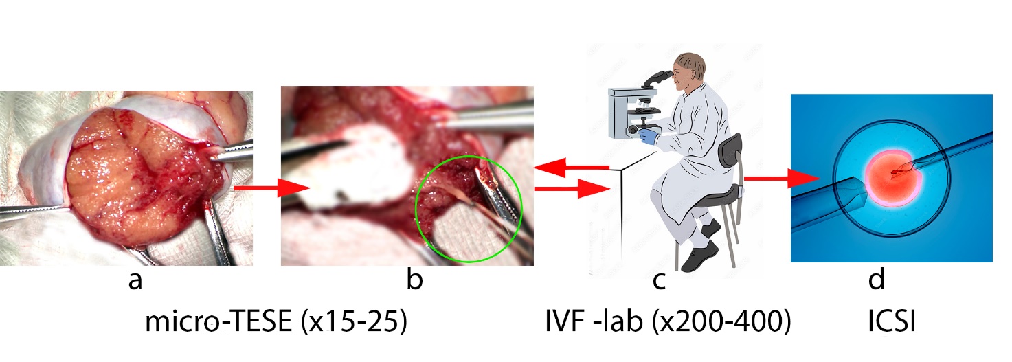
Figure 1:
Pipeline of the micro-TESE – IVF-ICSI cycle: a) revision of testicular tissue
using a surgical microscope, b) performing a biopsy from areas with the most
expanded tubules, c) examination of the resulting tissue in the IVF laboratory,
d) use of sperm for IVF- ICSI
It is important to note that the surgeon needs to examine a
relatively large area of testicular tissue in a limited operating time in order
to find a site suitable for biopsy. At the same time, the assessment of the
degree of maturity of the seminal tubules during micro-TESE is highly dependent
on the experience of the doctor, since it is based on a visual comparison of
the diameters of the tubules and the consistency of their contents. The method
of evaluation of testicular tissue, characterized by objectivity, high
specificity, harmlessness to reproductive cells, as well as the ability to
conduct analysis in real time and intraoperatively, can increase the
probability of detecting areas of spermatogenesis.
To interpret the laboratory data, it is desirable to have an
idea of the differences in testicular morphology in normal and in NOA [5]. So,
normally, testicular tissue consists of
tubuli seminiferi
and
interstitial tissue located between them
(interstitium)
(Fig. 2).
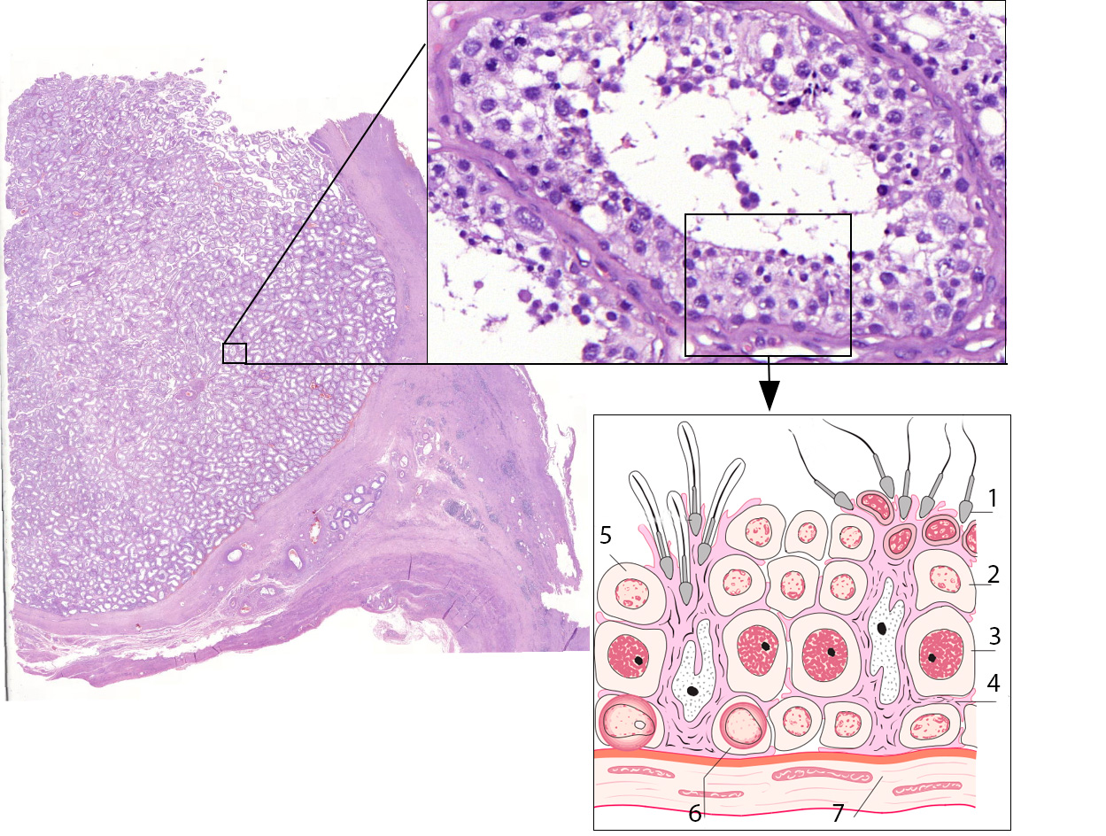
Figure 2:
Testicular tissue structure: 1 – spermatozoa, 2, 5 – round-cell spermatids, 3 –
spermatocytes, 4 – Sertoli cells, 6 – spermatogonia, 7 – peritubular cells
The tubular component – the vas deferens make up 60-80% of the
volume of the testicle. This is the place where the production of germ cells –
spermatozoa – is carried out. Each
tubulus semeniferi
has a wall
consisting of a layer of collagen and peritubular cells - myofibroblasts. The
lumen of the tubule is occupied by germinative cells, which differentiate into
spematozoa, and somatic cells, the main of which – Sertoli cells – have the
function of maintaining and regulating sperm maturation. All phases of germ
cell maturation are present in the tubule at the same time and are separated in
space – as spermatogenesis cells differentiate, they move from the periphery of
the vas deferens to its lumen, along the supporting Sertoli cells. Eventually,
mature spermatozoa are separated into the lumen and carried further along the vas
deferens. The interstitial component, which occupies 12-15% of the volume of
the testicle, is represented by loose connective tissue with nerve fibers,
blood and lymph vessels.
In the case of non-obstructive azoospermia, four histological
types can be detected simultaneously in the testicular tissue:
hypospermatogenesis – incomplete composition of the epithelium of the
tubuli
seminiferi; maturation arrest – if spermatogenesis stops at a certain
stage; Sertoli cells only syndrome (SCO)- only Sertoli cells are present,
germinative epithelium is absent and tubular hyalinization - Sertoli cells and
germinative epithelium is absent, the vas deferens are structurally
indistinguishable.
Advances in the development of IVF- ICSI today allow not only
mature sperm cells to be used for fertilization of an egg, but also their
precursors – round and late spermatids [6], which ultimately reduces the task
to differentiating the seminal tubules containing spermatogenesis cells from
those tubules where the spermatogenic epithelium is completely absent.
In order to improve the results of micro-TESE, various teams
of researchers are studying the possibility of using modern imaging methods,
which in the future would allow the surgeon to assess the structure of
testicular tissue with greater objectivity, increasing the probability of
detecting tubules containing sperm in the testicular tissue in NOA. These
methods can be divided into two groups. In the first case, physical
interactions of biological tissue and radiation are used that are inaccessible
to direct human observation, in the second – methods that improve the
visualization of the surgical field directly by the surgeon. The world
literature describes the experience of using a multiphoton microscope [7,8] to
study testicular tissue. Using a near-infrared laser source to induce
autofluorescence of tissues, this method creates a high-resolution image and
allows for high reliability (about 92%) in real time to distinguish seminal
tubules with normal spermatogenesis from pathological ones. However, the use of
laser radiation is potentially dangerous by thermal damage to DNA, which can
lead to further mutations. And although this laser has shown low phototoxicity
in rodent experiments, its safety for human DNA needs proof.
Another promising method is Raman spectroscopy [9,10]. Based
on the so-called raman scattering of photons, it is also a real-time
visualization method, with the help of which it is possible to detect seminal
tubules with preserved spermatogenesis with even greater accuracy (up to 96%)
in animal models with induced NOA-SCO. But the relatively long scanning time at
one point, which is about 2 minutes, limits the use of this method in a real
operation. All of the above is also true regarding the use of a laser as a
photon source.
Another
method
– full-field optical coherence tomography [11,12] – does not pose a danger
associated with the use of a laser, since a halogen lamp can serve as a light
source for it. The high speed of image acquisition - about 1 frame per second,
allows scanning a relatively large surface area of the tissue in a short time,
revealing the seminal tubules containing sperm by a characteristic reflected
signal from the microstructures of the sperm tails, which was also demonstrated
on an animal model. However, today this method has serious limitations for use
during micro-TESE due to insufficient resolution, low depth of tissue scanning
and difficulty in interpreting the results.
Another approach to solving the problem of improving the
quality of micro-TESE is to improve the visualization of the operating field.
The most modern microsurgery technology, the ORBEYE high-resolution 3D
microscope [13,14], creates a three-dimensional image of the surgical field
projected onto the surgeon's 3D glasses, and can improve the visualization of
testicular tissue, but tissue analysis remains subjective.
There are few works where computer image processing using a
neural network is used in order to improve the results of micro TESE. The
algorithm is based on the detection of tubules of the largest diameter, but
this approach has dubious advantages over conventional visual inspection [15].
Previous studies have established [16] that testicular tissue
has different optical properties depending on the degree of completion of
spermatogenesis. The use of spectral reflection characteristics as additional
data in the task of differentiation of testicular tissues can increase the
probability of detecting areas of spermatogenesis in NOA. The paper considers
the possibility of creating a specialized microsurgical system using the
spatial distribution of the spectral characteristics of testicular tissues to
assess its histological structure during micro-TESE. For this purpose, studies
of the reflection spectrum of testicular tissue with varying degrees of
spermatogenesis obtained during testicular biopsy in the visible and near
infrared ranges were carried out.
To simulate the reflection spectrum of testicular tissue
regions in NOA, testicular tissue samples were taken from patients who
underwent orchiectomy for various indications: severe testicular hypoplasia /
atrophy, trauma or inflammation of the testicle. The spectral characteristics
of tissues with preserved spermatogenesis were collected from biopsy material
obtained during autopsy in fertile men during their lifetime. A complete
absence of spermatogenesis was observed in the tissues of hard hypoplastic
testicles removed due to cryptorchidism. The selected tissue samples had a
relatively homogeneous structure, either with intact spermatogenesis or with
its complete absence, which excludes measurement errors associated with the
heterogeneity of the morphological structure of the samples.
After determining the histological structure of the obtained testicular
tissue (TT), its spectral characteristics were recorded by assembled setup
(Fig. 3) using spectrometers (S) OceanOptics FLAME-VIS and FLAME-NIR
(spectral range 350-1000 and 950-1650 nm, spectral bandwidth 1,34 and 10 nm,
exposure time range 3,8 ms–20 s and 1 ms–65 s, respectively). Reflected
radiation was introduced into the spectrometer using an optical fiber (OF) with
collimator (Cl) (field of view 5×5°) mounted at a fixed distance at an
angle of 45° to the sample plane. A halogen lamp Dedolight DLH4 (150 W) was
used as a light source (LS). Preprocessing of the spectrometer data
included Gaussian smoothing (
σ=
20) to eliminate high-frequency
noise. The reflection was extracted by normalizing the spectral brightness of
tissues to the spectral illumination created by the source in the sample plane.
To analyze the spectral features of healthy tissues and tissues with impaired
spermatogenesis, the spectral reflectance curves were normalized to the maximum
value and are presented in arbitrary units (a.u.). The spread of values was
determined as the minimum and maximum value of the reflection coefficient among
the obtained samples for each wavelength.

Figure 3:
Assembled setups
To determine the spatio-spectral tissues
characteristics, we assembled another setup consisting of a microscope (M) with
a 5x
magnification, a digital camera (C) with a wide spectral
sensitivity range TOUPCAM SWIR1300KMA (spectral sensitivity range 350-1700 nm,
pixel size 5×5 µm, exposure time range 50 µs-3600 s), custom nozzle with
a place for a filter (F) and a Dedolight DLH4 halogen lamp (150 W). The nozzle
was fixed in front of the camera sensor to enable registration of spectral
images. We used the Thorlabs FKB-VIS-10 and FKB-IR-10 filter sets, which allow
filtering in the range of 350-850 nm with a step of 50 nm from the visible
(VIS) to the near infrared (NIR) range and 900-1600 with a step of 100 nm from
the NIR to short-wave infrared (SWIR) range. The width of the spectral channels
of the filter sets is 10 nm. The microscope was focused on a tissue placed on a
microscope stage and pressed against a glass slide to eliminate glare.
The experimental protocol included adjusting a filter in a
special nozzle aperture, refocusing the microscope, and recording 50 spectral
images of the sample at the same exposure for each filter. Then, with the same
camera settings, spectral images of the reference plate were recorded with a
uniform reflection close to 1. By dividing the multispectral cube of tissue
images by the reference multicube, the spatial distribution of the spectral
reflectance of the samples was obtained. The spectral reflectance was then
averaged over the region and normalized to the maximum in the VIS-NIR and
NIR-SWIR region for comparison with the spectral characteristics obtained by
the fiber spectrometer. We also obtained correlation maps showing the degree of
correspondence between the pixels of the sample reflectance multicube and the
spectrum of healthy tissue.
The results of measuring the spectral
characteristics of healthy tissues and tissues with impaired spermatogenesis
are shown in the figure (Fig.4). Also, Fig. 4 shows the averaged
reflectance spectra over the areas of healthy samples, obtained using
multispectral imaging on a microscope. Discrete spectral data were interpolated
by a spline. Multispectral imaging data does not go beyond the spread of
reflectance values of a healthy tissue type, determined by the fiber
spectrometer.
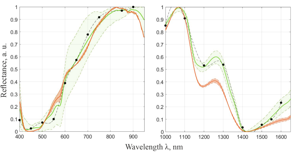
Figure 4:
The
average normalized spectral reflection coefficient of healthy tissues (green
curve) and tissues with NOA (red curve) with a corresponding spread of values
(colored areas) according to the samples. Black dots show averaged
multispectral data from a healthy sample
As a result of the correlation analysis of the multispectral
reflection cube of healthy tissue samples, maps were obtained (Fig. 5).
Comparing the correlation map with the image at 550 nm, it can be concluded
that the intertubular space and residual vignetting effects have a relatively
low correlation with the spectrum of healthy tissue, which will avoid false
negative classification. At the same time, most of the healthy tissue has a
uniformly high spatial correlation (above 0,99) with the spectrum of healthy
tissue.

Figure 5:
Tissue image (left) at 550 nm wavelength and
correlation map with average spectrum of healthy tissue (right)
The high correlation of the reflection
spectra of testicular tissues obtained from integral spectrometers and by
averaging images with filters, as well as the uniformity of the correlation map
constructed for healthy tissues, which corresponds to its morphological
uniformity, confirm the reliability of the obtained data. These data suggest
that in the infrared (IR) region of 1150-1400 nm there are noticeable
differences in the spectral characteristics of seminal tubules with preserved
and impaired spermatogenesis, which can be used for tissue differentiation in
clinical practice. In the visible region of the spectrum, the differences in
spectral characteristics are less pronounced and require further research and
accumulation of statistical material.
The identification of spectral features
characteristic of tissues with normal and impaired spermatogenesis is necessary
for the subsequent design of hardware for spectral differentiation of tissues.
Thus, the proposed surgical system can be built on the basis of multispectral
imaging and contain active illumination at different, predetermined wavelengths
or isolate the corresponding narrow spectral channels from broadband radiation
reflected from the studied tissues using light filters. The images recorded by
a monochrome video camera with high sensitivity and bitness in various channels
will contain the spatial distribution of the spectral characteristics of the
studied tissues and can be transmitted for processing and output to the
surgeon's monitor during the operation with markers in the areas with the
highest probability of the presence of spermatozoa. The safety of the proposed
method is determined by the use of incoherent radiation of LEDs or xenon lamps
used in modern surgical microscopes as light sources. We assume that the system
should be autonomous, not requiring a surgical microscope.
In this study, the possibility of creating a spectral method
of differentiation of testicular tissues with preserved and impaired
spermatogenesis in patients with NOA during IVF-ICSI was considered. We
proposed to use spectral characteristics as additional information about the
histological structure of testicular tissue, in addition to the traditionally
used diameter and consistency of the contents of the seminal tubules. To
confirm the possibility of creating a spectral method for searching for
spermatozoa in NOA, we implemented a number of
in vitro
experiments.
Reflection spectra of testicular tissue samples of patients with varying
degrees of spermatogenesis preservation were experimentally obtained by
assembled setup. We presented the spatial distribution of the tissue spectral
data, and showed differences between healthy tissue and tissue with impaired
germ cell production in the near IR range. The obtained experimental outcomes
can be useful for algorithms of visualization and automatic recognition of
damaged tissues. The described approach to the analysis of testicular tissue
can be non-contact, high-throughput, automated, safe for germ cells,
intraoperative and thus appears to be a promising diagnostic tool for clinical
practice.
This study received support from the Federal State Task Program by Scientific and Technological Center of Unique Instrumentation of the Russian Academy of Sciences (FFNS-2022-0010). This work was performed using the equipment of the Shared Research Facilities of the Scientific and Technological Centre of Unique Instrumentation of the Russian Academy of Sciences.
1.
Vahidi S. et al. Success rate and ART outcome of microsurgical
sperm extraction in non-obstructive azoospermia: A retrospective study,
International Journal of Reproductive BioMedicine. 2021. Vol. 19. № 9. P. 781
–
788.
2.
Wosnitzer, M.,
Goldstein, M., Hardy, M. P. Review of azoospermia, Spermatogenesis. 2014. Vol.
4. № 1. P. e28218.
3.
Schlegel P. N. Testicular
sperm extraction: microdissection improves sperm yield with minimal tissue
excision, Hum Reprod.
1999. Vol. 14. № 1. P. 131
–
135.
4.
Schlegel P. N., Sigman M., Collura B.,
et al.
Diagnosis
and treatment of infertility in men: AUA/ASRM guideline part I, J Urol. 2021.
Vol. 205. № 1. P. 36
–
43.
5.
E. Nieschlag, et al.
Male Reproductive Health and
Dysfunction 3rd Edition,
ISBN:
978-3-540-78354-1,
P. 11-20, 158-162.
6.
Goswami G, Singh S et al. Successful
fertilization and embryo development after spermatid injection: A hope for
nonobstructive azoospermic patients, J Hum Reprod Sci. 2015 Jul-Sep; 8(3): 175
–
177.
7.
Katz M.J., Huland D.M.,
Ramasamy R. Multiphoton microscopy: applications in urology and andrology,
Transl Androl Urol. 2014. Vol. 3. № 1. P. 77
–
83.
8.
Najari B.B., Ramasamy
R, Sterling J, et al. Pilot study of the correlation of multiphoton tomography
of ex vivo human testis with histology, J Urol. 2012. Vol. 188. № 2. P. 538
–
543.
9.
Huang W.E., Li M.,
Jarvis R.M., et al. Shining light on the microbial world: the application of
Raman microspectroscopy, Adv Appl Microbiol. 2010. Vol. 70. P. 153
–
186.
10.
Osterberg E.C., Laudano
M.A., Ramasamy R., et al. Identification of spermatogenesis in a rat
sertoli-cell only model using Raman spectroscopy: a feasibility study, J Urol.
2014. Vol. 192. № 2. 607
–
612.
11.
Ramasamy R., Sterling
J., Manzoor M., et al. Full field optical coherence tomography can identify
spermatogenesis in a rodent sertoli-cell only model, J Pathol Inform. 2012.
Vol. 3. № 1. P. 4.
12.
Jain M., Shukla N.,
Manzoor M., et al. Modified full-field optical coherence tomography: a novel
tool for rapid histology of tissues, J Pathol Inform. 201. Vol. 2, № 28. P.
82053.
13.
Best J.C., Gonzalez D.,
Alawamlh O.A., et al. Use of 4K3D video microscope in male infertility
microsurgery, Urol Video J. 2020. Vol. 7. № 1. P. 100046.
14.
Hayden R.P., Chen H., Goldstein M., et
al.
A
randomized controlled animal trial: efficacy of a 4K3D video microscope versus
an optical operating microscope for urologic microsurgery, Fertil Steril. 2019.
Vol. 112. № 3. P. E93.
15.
Pandya S., Halgrimson W., Pagani R.,
Finding
A Sperm Among the Weeds: Novel Neural Network Model for Augmented Seminiferous
Tubules Classification in MicroTESE. J Urol Vol. 201, No. 4S, Supplement,
Saturday, May 4, 2019.
16.
Yudovskii S.O., Kovylina M.V., Ryabova A.V. Combination of
autofluorescence diagnostics with the micro-tese for azoospermic patients,
International symposium on laser medical applications. Moscow. 2010.