The modern level of development of optical
electronics and computer technology allows creating and implementing the new
tools for study of biological objects. The method of interference microscopy is
one of the most promising. Itallows noninvasive studies of cell morphology and
dynamics with ultrahigh spatial resolution [1, 2]. Unlike many other microscopy
methods, the method of interference microscopy provides possibility of a native
object study without preliminary fixation and staining, [3]. The recorded value
of the optical difference in the interferometer allows obtaining quantitative
information on the volume distribution of the refractive index of the object
and its morphology which is an important feature of interference methods [4].
Estimation of the phase height distribution reconstructs a true
three-dimensional surface of the object [5]. In our opinion, a study of optical
density and reproduction of cell geometry provides additional information about
blood cells, which are traditionally studied by methods of light microscopy.
The aim of the work is to discuss and demonstrate
the new methodological approaches to the analysis of neutrophils and
erythrocytes state in norm and under stress using interference microscopy.
The method of interference microscopy is based on
the principle of measuring phases which are normalized quantities and determine
the optical parameters of the object. The method of phase steps at control of
polarization registers phase images of object [3]. The phase difference of the
object is calculated by the phase-step method and is schematically represented
as follows:
Where I0-3(x,y)
– the distribution of radiation intensity in the field of view of the
photodetector, k – the wave number, d – the shift value of the reference
mirror.
The required value of the phase difference is
determined by taking into account the intensity distribution in the field of
view of the photodetector:
Where I3(t0) – the
instantaneous value of intensity determined by the exposure time of the
photodetector.
Interference images of 1280x1024 pixels include 3
frames per second and 128x128 pixels up to 250 frames per second. This variant
of phase step counting in phase images achieves spatial superresolution [6]
which gives additional information compared to classical images obtained by
light microscopy. The advantages of measuring phase steps using interference
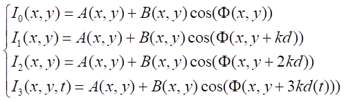
microscopy can be clearly demonstrated by Figure 1.
Figure 1 shows the interpretation of the phase
step measurement, taking into account the individual pixels.
Figure 1 – Measuring
phase steps using interference microscopy
The solid line in Figure 1 (a) shows the
interferogram obtained for the pixel highlighted in red with coordinates (1,1).
The interferogram in this case represents the time dependence of the radiation
intensity value in the field of view of one pixel of the photodetector and
reflects the sequence of single-pixel fragments of thousands of 1024x1024 pixel
interferograms. Then, as highlighted with a dotted line n in Figure 1(a)we
obtain the dependence of the radiation intensity for the adjacent (blue) pixel
with coordinates (1, 2) and calculate the phase value for this area using the
relation δφ=φ(1, 2)-φ (1, 1). We obtain a complete phase
image of the object by repeating this sequence for all pixels.
Thus high spatial resolution
of the microscope is determined by decrease of phase steps (Figure 1a, 1b),
whereas with increase of phase steps the confidence interval from ∆1 to
∆2 of phase value in different points of sinusoid is calculated rather
roughly (Figure 1c). As a result of the construction of interferograms the
resolution of interference microscopy reaches 0.1 nm in the vertical and 15 -
100 nm in the plane of the object.
The optical scheme of the laser channel is a
modification of the Mach-Zehnder interferometer based on independent
polarization control in the object and reference arms of the interferometer.
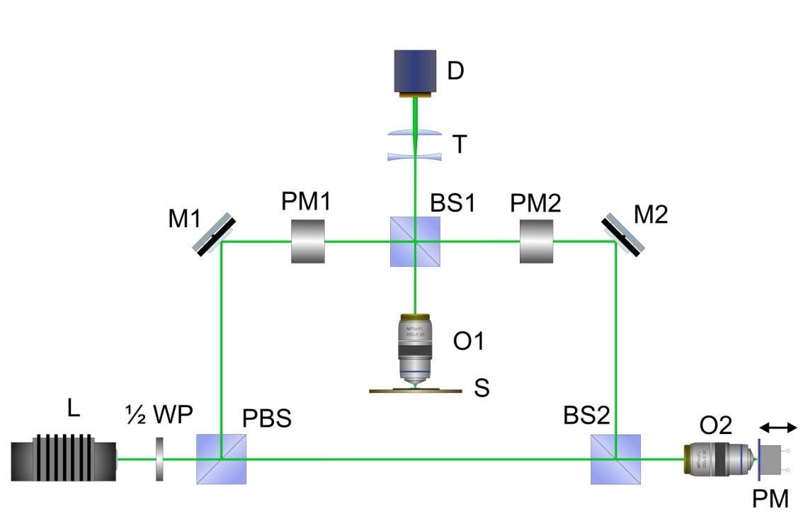
Figure 2 – Optical scheme
of the laser channel of the interference microscope.
As shown in Figure 2 the laser beam from the
laser (L) is divided into two at the polarizing beam splitter PBS. One beam
(objective) is focused by the objective O1 on the object which is placed on the
stage S and reflecting from the mirror substrate through the light splitter BS1
and the telescopic system T gets to the photodetector D - camera Silicon
Imaging model SI - 1280f. Another beam (reference) does not pass through the
object and is focused by the lens O2 and reflected from the mirror of the piezoelectric
transducer (PM) and the same falls into the photodetector, where the
interference of beams occurs and a phase image is formed. At construction of
the phase image the signal is normalized by wavelength and the optical
difference of a course of beams which characterizes value of phase height of
object (F) in the given point is determined:
Where ȹ0
– the initial phase,
ȹ0bj
– the phase shift by the object, λ – the wavelength
of radiation, Φ0
– the constant shift which is determined by
the choice of the initial phase reference point.
Registration of phase height (Φ) in all
points of the object forms 3D images. Processing of the received data is
carried out with LabView software. Dynamic phase images are obtained and
processed in the Airyscan V.6 software. The user interface of Airyscan V.6
consists of two parts: the panel of parameters determining correct operation of
the program (the left part) and the main elements of the Tab Control (the right
part) which display phase images (Figure 3).
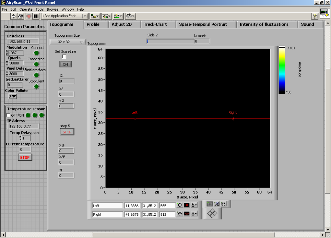
Figure 3 – Airyscan
program interface
Topo3D software is used to playback 3D images.
The program sequentially reproduces a series of static phase images and
displays a 3D image with a possible cross section of phase images.
Figure 4 – Toro3D program
interface
Playback modes include a phase image playback
page with the ability to crop images using two cursors that define the clipping
area, allowing you to maintain the dimensionality of the clipped images.
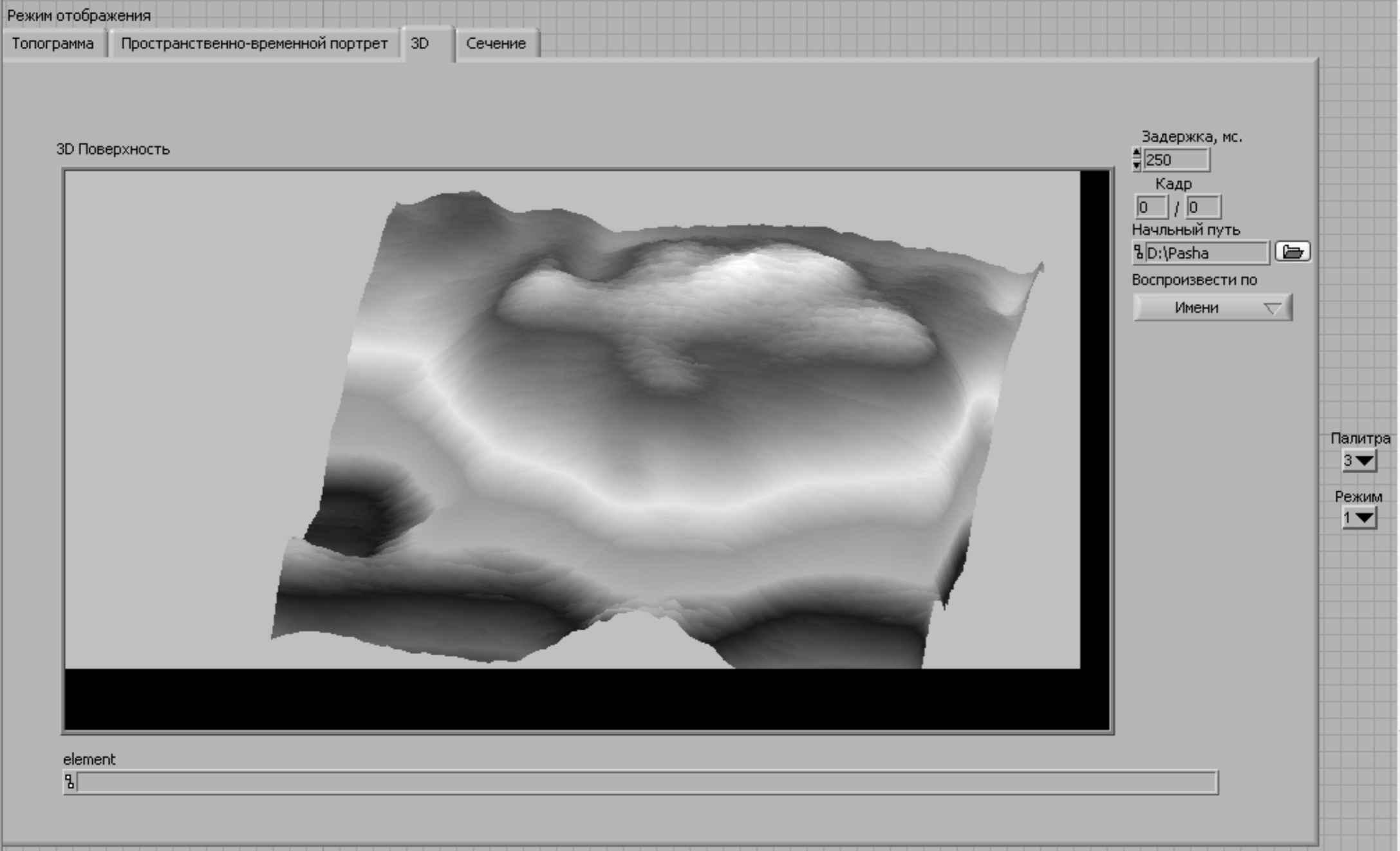
Figure 5 – 3D phase
images mode window
Analysis of neutrophils and erythrocytes under
technological stress was carried out by interference microscopy with further 3D
reconstruction according to the purpose of the study. 30 clinically healthy
high-yielding black and gray cows were the object of study. The choice of the
research object was dictated by the strategic task of modern animal breeding
associated with the reduction of losses caused by technological stress.
Technological stress causes higher susceptibility of animals to pathogens and
reduction of animal productivity. A combination of factors: regrouping, change
of service personnel and veterinary and sanitary manipulations were
technological stress for animals. During the study, blood sampling in all
experimental animals was carried out before the technological stress and after
the action of the combination of factors causing technological stress.
Neutrophils were isolated by the standard method
on a double density gradient (1.077 and 1.093). The cells were washed by
centrifugation in Hanks's solution. The supernatant was poured off, and
neutrophils were diluted with Hanks' solution to a concentration of 2x10⁶.
Erythrocytes were examined in whole blood.
The morphofunctional state of cells was assessed
by computer phase morphometry based on a domestic computer laser
phase-interference microscope MIM-340 (Yekaterinburg, Russia) [7].
Leukocytes and erythrocytes were additionally
examined in smears stained by Romanovsky-Giemsa. The morphology of leukocytes
and erythrocytes was examined on a light microscope Micromed C-11 (Russia) with
MECOS-C software.
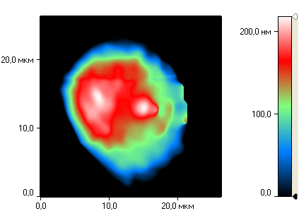
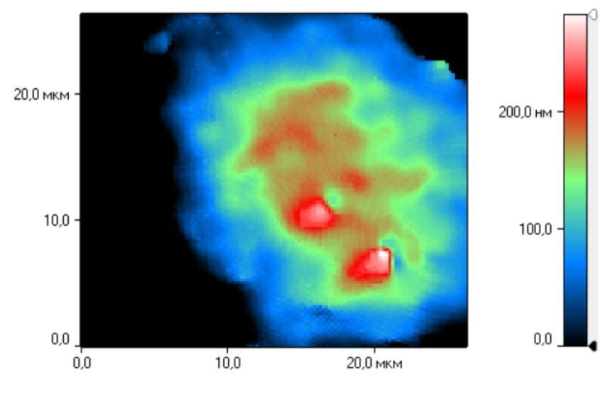
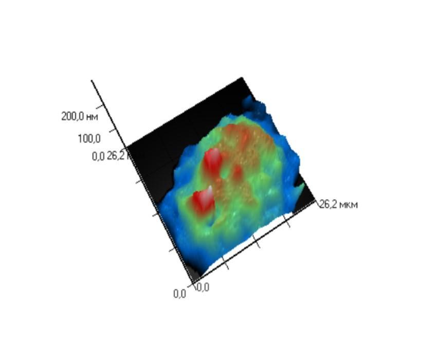
Interference microscopy of neutrophils in intact
animals not subjected to technological stress allows us to identify the two
most distinct cell populations (Figure 6).
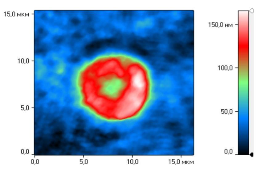
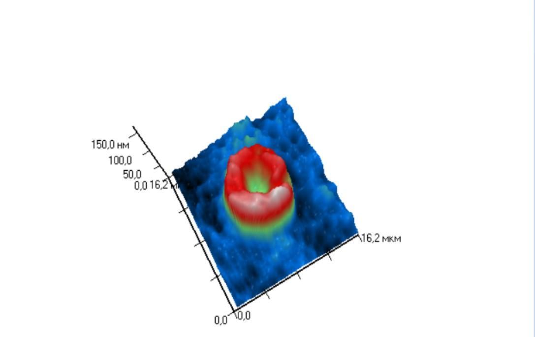
Figure 6 – Different
morphological types of neutrophils. Phase image (topogram) (A) and 3D
reconstruction of cell image (B) of morphological type I. Phase image
(topogram) (C) and 3D reconstruction of cell image (D) of morphological type
II.
The first population of neutrophils is
represented by the Ist morphological cell type [8]: 3D reconstruction of the
cell image clearly demonstrates that these are round-shaped cells with a
clearly distinguished nucleus and uniform distribution of intracellular contents
(Figure 6 A, B). 3D reconstruction of the second cell population displays an
uneven surface with many convexities and depressions. This is due to the
spatial redistribution of the cytoplasm, intracellular organelles and nucleus.
This is morphological type II (Figure 6 C, D).
Counting the I and II morphological cell types
showed that under technological stress, the I morphological type decreased by 3
times and the II morphological type of neutrophils increased by 2 times.
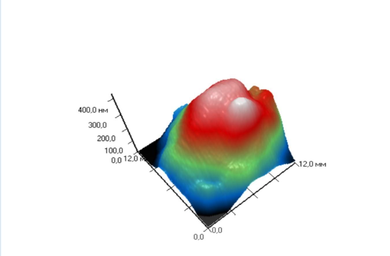
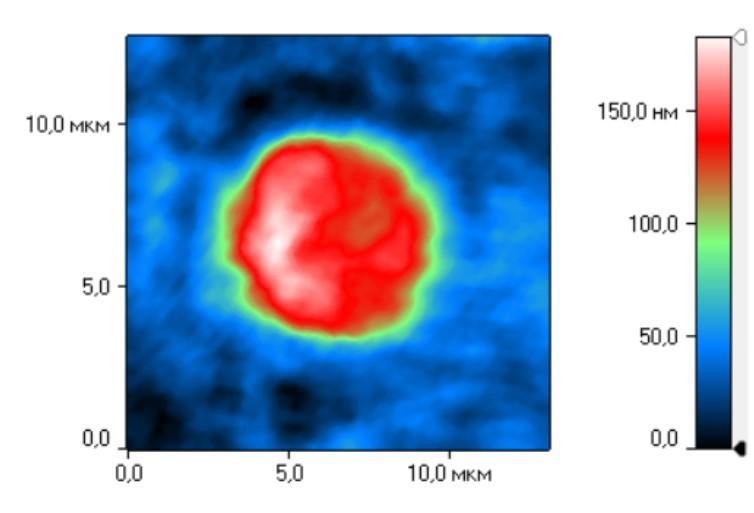
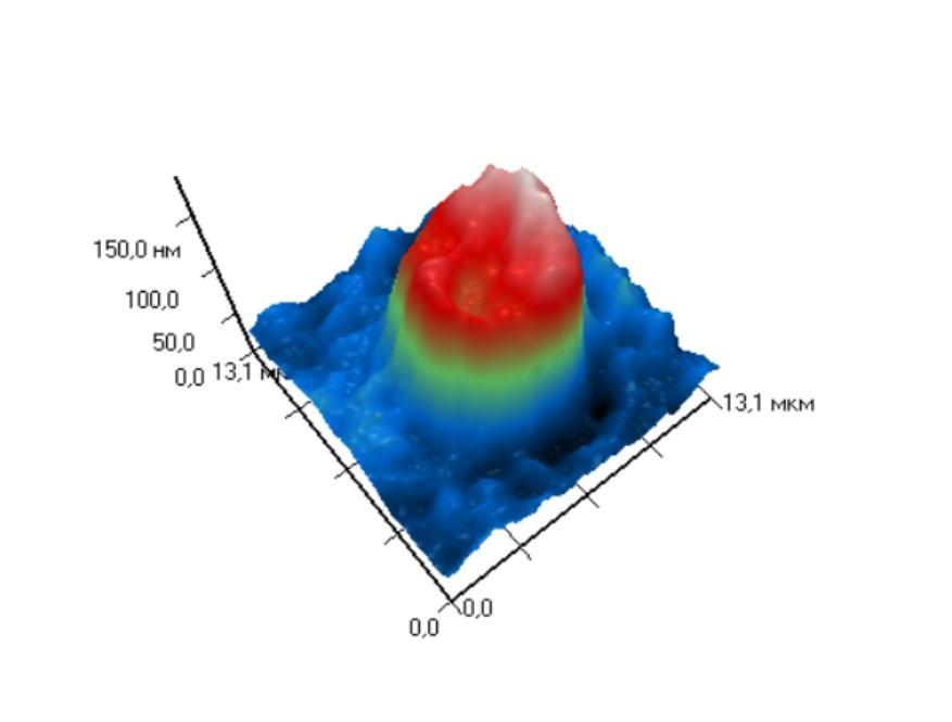
Analysis of erythrocyte morphology by
interference laser microscopy revealed that before technological stress,
erythrocytes were characterized by a normal biconvex cell shape (Figure 7 A,
B). Technological stress caused a change in the shape of the erythrocytes. 3D
reconstruction of the cell images revealed spikes, ridges, and protrusions on
the cell surface (Figure 7 C, D).
Figure 7 – Different
morphological forms of erythrocytes. Phase image (topogram) (A) and 3D
reconstruction of cell images (B) of erythrocytes before technological stress.
Phase image (topogram) (C) and 3D reconstruction of cellular images (D) of
erythrocytes after stress.
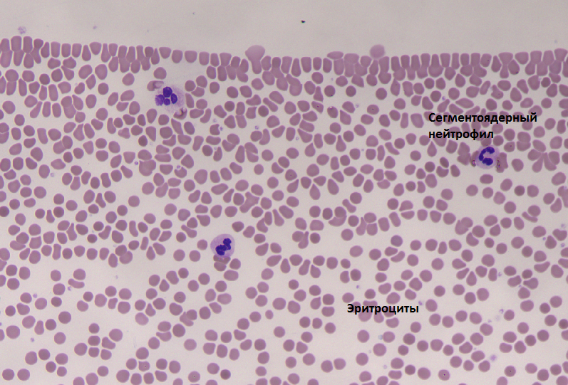
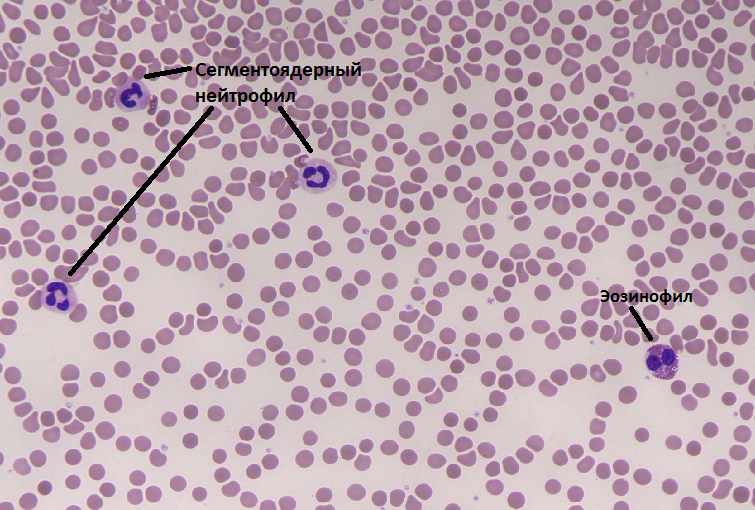
It should be noted that unlike 3D reconstruction
of cell images, the use of light microscopy does not provide an opportunity to
determine the change in the shape of neutrophils and erythrocytes (Figure 8).
All cells have a rounded shape.
|
|
|
|
A
|
B
|
Figure 8 – Cell
morphology under the light microscope.
A – before stress, B –
after stress
Based on the results obtained, we can conclude
that the reconstruction of 3D images allows cell morphology analysis and
significantly complements representation obtained with the light microscopy
data. In addition, imaging is the basis for the analysis of the mechanism of
stress exposure.
Neutrophils are the first protective cell barrier
against infections, the most numerous phagocytes in the human body which are
quickly mobilized from the bloodstream to the infectious focus or site of
injury [9]. Deformed cell contours indicate a certain degree of cell activity
[10]. An increase in morphologically altered erythrocytes is of key importance.
Changes in erythrocyte morphology are reflected in the oxygen-transporting
function of blood [11] which leads to impaired blood supply to tissues [12].
The study reveals the possibility of using laser
interference microscopy to assess cell morphology within nanometer scale range.
Analysis of the results reveals changes in the cellular link of nonspecific
resistance and deterioration of the oxygen transport function of erythrocytes
under technological stress.
3D imaging
using interference microscopy allows us to quickly identify and obtain the most
informative data on the geometric parameters of cells. Blood cells in this
method of analysis are not subjected to additional sample preparation before
the study (fixation, staining, treatment with contrasting agents), which
minimizes the possibility of artifacts. In addition, 3D computer images are
lifetime visualization of cellular processes and can be obtained in a very
short time. The mentioned facts represent the most important condition for
further development of works in the field of cell diagnostics. Using computer
methods of cytodiagnostics in the work revealed new aspects of functional
morphology of neutrophils and erythrocytes under stress. The obtained data are
of fundamental importance for the development of new methods for rapid
diagnosis of the adaptation reserve at the cellular level.
This work was supported by Grant № 22-26-00311 of
the Russian Science Foundation.
[1] Lue N., Popescu G., Ikeda T., Dasari R.,
Badizadegan K., Feld M. Live cell refractometry using microfluidic devices //
Opt. Lett., 2006, 31(18), Р. 2759-2761
doi: 10.1364/ol.31.002759
[2] Park Y., Popescu G., Badizadegan K., Dasari
R., Feld M. Diffraction phase and fluorescence microscopy // Opt. Expr. 2006,
14(18), Р.8263-8268.
doi: 10.1364/oe.14.008263
[3] Vishnyakov G.N., Levin G.G, Minaev V.L.,
Pickalov V.V., Likhachev A.V. Tomographic interference microscopy of living
cells // Microscopy and Analysis, 2004, № 87, Р. 19-23.
[4] Zagubizhenko M. V., Yusipovich A. I., Pirutin
S. K., Minaev V. L., Kudryashov Yu. B. Use of laser interference microscopy to
study the state of mouse peritoneal macrophages irradiated with ultraviolet
light // Radiation Biology. Radioecology, 2011,V. 51. № 6, P. 715.
[in Russian].
[5] Deryugina A.V., Ivashchenko M.N., Ignatiev P.S.,
Lodyanoy M.S., Samodelkin A.G.
Alterations in the phase
portrait and electrophoretic mobility of erythrocytes in various diseases
//
Modern
Technologies in Medicine,
2019, Т. 11.
№ 2,
С. 63-68.
doi:
10.17691/stm2019.11.2.09.
[6] Tychinsky V.P., Kretushev A.V., Vyshenskaya
T. V., Tikhonov A.N. Coherent phase microscopy in cell biology: visualization
of metabolic states // Biochim. Biophys. Acta,
2005, № 1708, Р. 362-366.
doi: 10.1016/j.bbabio.2005.04.002
[7] Deryugina A.V., Ivashchenko M.N., Ignatyev
P.S., Belov A.A., Petrov V.A. Diagnostics possibilities of erythrocytes
analysis by the method of laser interference microscopy // Klinicheskaya
Laboratornaya Diagnostika, 2021, V. 66, № 1. P. 22-25. doi:
10.18821/0869-2084-2021-66-1-22-25
[8] Vasilenko I.A., Nikitin A.A., Malichenko
N.V., Ivanjuta I.A., Metelin V.B., Agadzhanyan B.J. Сytometry of neutrophils
for evaluation of complex treatment efficiency in patients with mandibular
osteomyelitis // Clinical Medicine Almanac, 2008, №.18, Р. 63-68. [in Russian].
[9] Mantovani A., Cassatella M.A., Costantini C.,
Jaillon S. Neutro-phils in the activation and regulation of innate and adaptive
immunity // Nat. Rev. Immunol, 2011, № 11, Р. 519-531. doi: 10.1038/nri3024
[10] Tuzlukov I.I. Features of the morphology of
neutrophilic leukocytes in the postpartum period // Pavlov Russian Medical and
Biological Bulletin, 2004, № 3-4, P. 113-117. [in Russian].
[11]
Kobayashi S.D., Malachowa N., DeLeo
F.R. Influence of microbes on neutrophil life and death // Front. Cell. Infect.
Microbiol, 2017, № 7, Р. 159. doi: 10.3389/fcimb.2017.00159
[12] Emelianov V.V., Leontev D.V., Ishchenko
A.V., Bulavintseva T.S., Savateeva E.A., Danilova I.G.. Atomic force microscopy
imaging of red blood cells and metabolic disorders in experimental diabetes
mellitus and its correction with lipoic acid // Biophysics, 2016, V. 61. № 5,
P. 922-926. [in Russian].